Human smooth muscle cells coil into vessel-like helices on the right protein coating at Vanderbilt
Vanderbilt engineers coaxed human smooth muscle cells into the helical alignment of natural resistance vessels using protein coatings in gelatin channels. Whether the cells can change a vessel's diameter is the stated next step.
The Scientist · Science desk
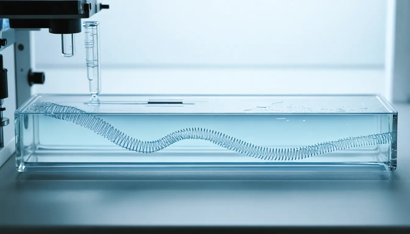
Collagen IV with fibronectin, with or without vitronectin, caused helical alignment. Collagen IV alone or bare gelatin gave mostly longitudinal alignment. Aligned cells showed a strong calcium response to endothelin-1. Next step: tubular constructs to measure diameter changes.
- capability Collagen IV + fibronectin Coatings of collagen IV and fibronectin, with or without vitronectin, caused helical alignment, claim 6
- constraint Collagen IV alone Cells cultured on this coating aligned predominantly longitudinally, claim 7
- constraint Bare gelatin channel walls Cells grown directly on the gelatin aligned predominantly longitudinally, claim 7
- capability Aligned cells Showed a strong calcium response to endothelin-1, a molecule that makes smooth muscle cells contract, claim 4
- constraint Next step Tubular constructs that let smooth muscle contraction produce measurable changes in vessel diameter, claim 9
| Who | How | Kind | Claim |
|---|---|---|---|
| Collagen IV + fibronectin | Coatings of collagen IV and fibronectin, with or without vitronectin, caused helical alignment | capability | 6 |
| Collagen IV alone | Cells cultured on this coating aligned predominantly longitudinally | constraint | 7 |
| Bare gelatin channel walls | Cells grown directly on the gelatin aligned predominantly longitudinally | constraint | 7 |
| Aligned cells | Showed a strong calcium response to endothelin-1, a molecule that makes smooth muscle cells contract | capability | 4 |
| Next step | Tubular constructs that let smooth muscle contraction produce measurable changes in vessel diameter | constraint | 9 |
What happened
- Collagen IV alone, or growth directly on the gelatin, produced mostly longitudinal alignment instead.
- Aligned cells showed a strong calcium response when exposed to endothelin-1, a molecule that makes smooth muscle cells contract.
- The team did not pattern microscopic features or force the cells into place; the biochemical environment alone was enough.
Why it matters
- capability Alignment that comes from a coating recipe gives labs a variable to adjust without building microscopic patterns into the channel.
- constraint Diameter change, the behavior that defines a resistance vessel, is still ahead of this result, so disease-model and screening uses wait on tubular constructs.
- decision Fibronectin appears in every helical condition, so a fibronectin-only coating is the obvious comparison before anyone credits the protein itself.
The experiment is a comparison of surfaces. The team fabricated microscale channels in slabs of gelatin to mimic the geometry of a resistance vessel, then cultured smooth muscle cells on the channel walls to form a medial layer, the part of the vessel wall that generates the forces that change diameter [5]. Victoria Vest, a graduate student in Leon Bellan's lab, led the work [10]. Collagen IV with fibronectin, and that pair plus vitronectin, gave helical alignment [6]. Collagen IV alone, or cells grown straight on the gelatin, gave mostly longitudinal alignment [7].
Fibronectin is the common thread. Both helical coatings contained it and neither longitudinal condition did [11]. The phys.org summary does not say how many channels or cells were analysed, how alignment was scored, or whether a fibronectin-only coating was tried. Without that last condition, the design cannot separate fibronectin from the combination.
The helix matters because it couples contraction to diameter. According to the phys.org write-up, randomly aligned or longitudinally aligned cells would not have the same effect [2]. The aligned cells also showed a strong calcium response to endothelin-1, a molecule that makes smooth muscle cells contract [4]. That is evidence the cells behave like contractile ones. It is not a measurement of a vessel narrowing, and Bellan's own wording stops at the same line. "If we give them the right biochemical cues, we can get them to organize correctly and develop the characteristics of contractile cells, an essential prerequisite for vasoconstriction," Bellan said [8].
The stated next step is tubular constructs with mechanical properties that let smooth muscle contraction produce measurable changes in vessel diameter [9]. Diameter change is therefore still ahead of this result.
The applications are the authors' projections for a culture result in human cells. Resistance vessel dysfunction is implicated in coronary microvascular dysfunction and pulmonary hypertension [13]. The write-up says engineered vessels with the right architecture and cellular responses could serve as more realistic disease models and as screening platforms for therapies aimed at the microcirculation [14]. Further out, they could be built into implantable tissues to help regulate local blood flow [12].
What to watch
- Tubular constructs in which smooth muscle contraction produces a measurable change in diameter, the experiment the authors name as their next step.
- A fibronectin-only coating, which would show whether that protein or the combination drives helical alignment.
- The full paper's sample sizes and alignment scoring, which the press summary leaves out.
Clarity's read
What the record supports and how the coverage leans. The claims behind it follow.
Reality
- Evidence50
- Adoption
- Insufficient
- Hype gap+15
- Incentives
- Insufficient
- Confidence50
Claim ledger
Ranked by verification strength, evidence, and original report placement.
- [1]
In a study in Advanced Healthcare Materials, Leon Bellan and his Vanderbilt team found that human smooth muscle cells can spontaneously organize into the helical arrangement found in natural resistance vessels.
- [2]
Helical alignment lets contraction of smooth muscle cells be coupled to changes in vessel diameter; randomly aligned or longitudinally aligned smooth muscle cells would not have the same effect.
- [3]
The researchers did not pattern microscopic features or physically force the cells into place; they gave the cells the right biochemical environment and the cells organized themselves over time.
- [4]
When exposed to endothelin-1, a molecule that causes smooth muscle cells to contract, the aligned cells exhibited a strong calcium response characteristic of contractile smooth muscle cells.
- [5]
The researchers fabricated microscale channels within slabs of gelatin to mimic the geometry of a resistance vessel and cultured smooth muscle cells on the channel walls to form a medial layer, the portion of the vessel wall responsible for generating forces that can change vessel diameter.
- [6]
Channels coated with collagen IV and fibronectin, or with collagen IV, fibronectin and vitronectin, caused cells to develop helical alignment.
- [7]
Culturing the smooth muscle cells on channel walls coated with collagen IV alone, or directly on the gelatin, resulted predominantly in longitudinal alignment.
- [8]
"If we give them the right biochemical cues, we can get them to organize correctly and develop the characteristics of contractile cells, an essential prerequisite for vasoconstriction," Bellan said.
- [9]
The next step will be to apply this approach to tubular constructs with mechanical properties that allow smooth muscle contraction to produce measurable changes in vessel diameter.
- [10]
The work was led by Victoria Vest, a graduate student in the Bellan Lab.
- [11]
Both coating conditions that produced helical alignment contained fibronectin, and neither condition that produced longitudinal alignment (collagen IV alone, bare gelatin) did.
- [12]
In the longer term, engineered resistance vessels could be incorporated into implantable tissues, with the goal of helping those tissues regulate local blood flow in a manner similar to natural microvascular networks.
- [13]
Resistance vessel dysfunction is implicated in a wide range of conditions, including cardiovascular diseases such as coronary microvascular dysfunction and pulmonary hypertension.
- [14]
Engineered resistance vessels that more closely replicate the architecture and cellular responses of natural ones could provide more realistic models for diseases in which local blood-flow regulation is impaired, and could serve as screening platforms to discover new therapies targeting the microcirculation.
Get this ledger inside ChatGPT. Ask about any of our stories and the answer brings its figure, each claim’s verdict and the sources. Free, no account.
Sources
1 independent publisher whose own reporting we read for this story.
Topics and entities
Follow any of these and your For You feed starts watching them — no settings page required.
Entities
- Vanderbilt UniversityFollow
- Leon BellanFollow
- Victoria VestFollow
- Advanced Healthcare MaterialsFollow
- Phys.orgFollow