Science1 publisherNot yet confirmed elsewhere2 min readPublished
Tampere lensless imager recovers quantitative phase from a single frame inside a cell incubator
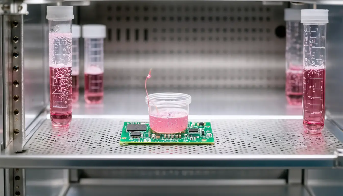
Tampere University's SF-PULSE recovers quantitative phase images of unstained living cells from a single frame, in a system small enough to sit in an incubator. According to the university, labs can then follow individual cells for hours or days without taking the dish out.
The Scientist · Science desk
What happened
- SF-PULSE combines an AI-based neural network with physics-informed image reconstruction to recover the phase signal.
- Recovering phase in lensless systems has traditionally required several images or added optical components, the computational problem the team set out to remove.
- Conventional quantitative phase imaging relies on large, complex microscopes that are poorly suited to long-term work inside a cell-culture incubator.
- The university says the compact system's wide field of view can cover hundreds or even thousands of cells at the same time.
Compiled by The ScientistSomething wrong?How this is made
Why it matters
- decision Labs running time-lapse studies could stop carrying dishes to a bench microscope, so the length of an experiment would no longer be capped by imaging sessions.
- cost Dropping extra exposures and optics moves the effort into computation. The cost of reconstructing each frame will decide whether days of continuous imaging can run on modest hardware next to the incubator.
- constraint The release gives no baseline or effect size, so labs cannot yet compare SF-PULSE's phase accuracy with the bench phase microscopes it would replace.
The paper appeared in Applied Physics Letters [4]. Its title is more specific than the press release about how the job is divided: "Single-frame lensless phase retrieval using learned sensor plane initialization" [5]. Taken at its word, the network supplies a starting estimate at the sensor plane, and the physics-informed reconstruction produces the final image [2]. I think that split is sensible. A network asked only for a starting point leaves the answer to an optical model, and an optical model is easier to audit than a network asked to output the phase map directly.
Igor Shevkunov, the study's first author, is from Tampere's Faculty of Information Technology and Communication Sciences [9]. He said: "Our SF-PULSE method can reconstruct quantitative phase information from a single measurement image without the need for complex optical hardware. The method reduced computational errors, improved reconstruction stability and enabled accurate long-term tracking of individual cells." [12] The first two gains are comparisons. The thing this doesn't tell you is what they were compared against or how large they were. It also leaves out which cells were imaged and how long one reconstruction takes.
Quantitative phase imaging measures growth, size and changes in biomass in transparent cells, with no labels [6]. Meenakshisundaram Kandhavelu is a university lecturer who heads the Molecular Signaling research group in Tampere's Faculty of Medicine and Health Technology [11]. He said the value lies with single cells: "Importantly, researchers can follow not only how a cell population changes, but also how individual cells grow, move, divide and gain or lose biomass over time. This is often more informative than examining the average behavior of a cell population alone." [10]
The university lists drug-response studies, toxicity screening and investigations of cell-death mechanisms as work the method could support [14]. All three depend on per-cell biomass readings staying accurate over days of continuous imaging. Before treating SF-PULSE as a screening instrument, I'd want to see that accuracy measured against a reference microscope imaging the same cells.
What to watch
- The full Applied Physics Letters paper's phase-error figures against a multi-frame or interferometric reference, and whether they hold across multi-day runs.
- A biological study from Kandhavelu's Molecular Signaling group that uses SF-PULSE for drug-response or toxicity work.
- A reported reconstruction time per frame, and the computing hardware needed to sustain it.
Clarity's read
What the record supports and how the coverage leans. The claims behind it follow.
Reality
- Evidence35
- Adoption
- Insufficient
- Hype gap+30
- Incentives35
- Confidence40
Claim ledger
Ranked by verification strength, evidence, and original report placement.
- [1]
In a study led by Tampere University, researchers developed a lensless imaging method, SF-PULSE, that produces quantitative data on living cells from a single image.
- [2]
SF-PULSE is a lensless quantitative phase imaging method that combines an AI-based neural network with physics-informed image reconstruction.
- [3]
The compact system can operate inside a standard cell-culture incubator, allowing cells to be monitored continuously for hours or even days without staining or repeated removal from the incubator for imaging.
- [4]
The research is published in the journal Applied Physics Letters.
- [5]
The paper is Igor Shevkunov et al, 'Single-frame lensless phase retrieval using learned sensor plane initialization', Applied Physics Letters (2026).
- [6]
Quantitative phase imaging enables observation of living, transparent cells without staining and provides quantitative information about cell growth, size and changes in biomass.
- [7]
Conventional quantitative phase imaging methods typically rely on large and complex microscope systems that are not well suited for long-term monitoring inside a cell-culture incubator.
- [8]
Recovering phase information in lensless systems is a challenging computational problem that has traditionally required multiple images or additional optical components.
- [9]
Igor Shevkunov is the study's first author, from the Faculty of Information Technology and Communication Sciences at Tampere University.
- [10]
"Importantly, researchers can follow not only how a cell population changes, but also how individual cells grow, move, divide and gain or lose biomass over time. This is often more informative than examining the average behavior of a cell population alone."
- [11]
Meenakshisundaram Kandhavelu is a university lecturer in the Faculty of Medicine and Health Technology at Tampere University and head of the Molecular Signaling research group.
- [12]
"Our SF-PULSE method can reconstruct quantitative phase information from a single measurement image without the need for complex optical hardware. The method reduced computational errors, improved reconstruction stability and enabled accurate long-term tracking of individual cells."
- [13]
According to the release, the compact lensless system can monitor hundreds or even thousands of cells simultaneously across a wide field of view.
- [14]
According to the release, the method could support applications such as drug-response studies, toxicity screening and investigations of cell-death mechanisms.
Sources
1 independent publisher whose own reporting we read for this story.
- phys.orgLensless imaging method enables long-term monitoring of living cells without staining
1 article · October 8, 2026
Topics and entities
Follow any of these and your For You feed starts watching them — no settings page required.
Topics
- Lensless imagingFollow
- Physics-Informed Machine LearningFollow
- Quantitative phase imagingFollow
- Live-cell imagingFollow