ScienceNot yet confirmed elsewhere1 publisher3 min readPublished
Atomic force microscopy shows two DNA helices zipped together groove to groove
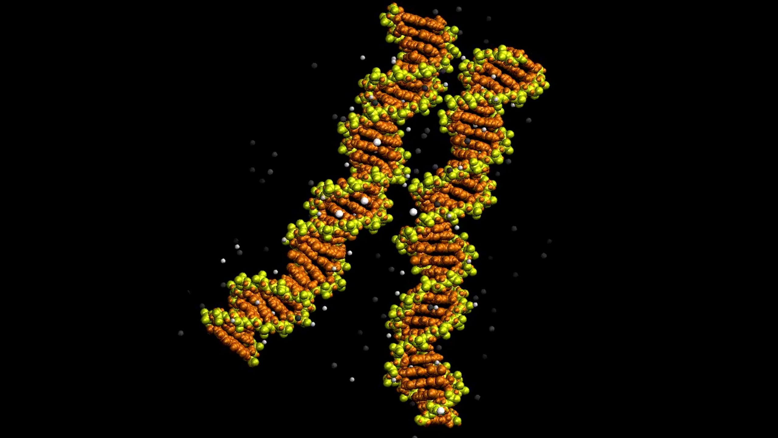
Sheffield and York researchers imaged two DNA helices with grooves aligned like a zip, confirming a pairing predicted more than 20 years ago. Simulations credit bridges of positive ions that form most readily at certain sequences, a model so far tested only on purified DNA.
The Scientist · Science desk
What happened
- In atom-level simulations, ions carrying two positive charges bridged neighbouring helices, with some of the strongest contacts where minor grooves lined up.
- With nickel ions the most stable simulated contacts sat at the short sequence GTAC, while magnesium and calcium formed different bridge networks.
- Most pairing arrangements seen under the microscope could not have come from fully matching sequences aligning along the whole contact.
Why it matters
- capability Because the model ties easy pairing to specific sequences such as GTAC, it can be tested by changing the sequence and measuring whether pairing gets easier or harder.
- constraint Models of how matching DNA finds its partner now have to treat ion-bridged first contact and sequence-matched extension as separate steps, since contact alone does not signal a match.
- constraint Conclusions about how genomes pack in a nucleus have to wait until the effect is shown with histones and other proteins present, because this work used purified DNA.
Seeing two DNA molecules touch would not settle anything [19]. The zipper model makes a specific geometric claim: the grooves spiralling around one helix line up with those of its neighbour. So the images had to resolve individual grooves [19][7]. That requirement shaped the method. Each molecule was already a double helix, and the team ran atomic force microscopy in liquid to see how two of them sit alongside each other [20][7]. The groove-level images were taken with nickel ions because nickel gave the best spatial resolution. Magnesium and calcium appeared in larger-area scans and in simulations [8].
"The microscopy images were taken in static form, which allows us to obtain the resolution where we can observe and measure the individual minor and major grooves on each molecule," Sheffield biophysicist Alice Pyne told ScienceAlert [9]. The movie of DNA progressively zipping together comes from simulations, not from the microscope [17].
"The way two DNA duplexes zip together was hypothesized over 20 years ago," York biophysicist Agnes Noy told ScienceAlert. "These images represent the first visualization that this idea is real." [10]
The harder question is why two molecules carrying the same negative charge stay together at all [6]. "The ions help create a salt bridge between the two molecules, which holds them together," Pyne said [11]. In simulations that tracked individual atoms, ions carrying two positive charges spanned the gap between neighbouring helices. Some of the strongest contacts formed where the minor grooves aligned [12].
Sequence mattered in those simulations. "What the simulation showed is that there are special sequences that preferentially form these bridges, pinning the two molecules together, which then allows the molecules to 'zip' together," Pyne said [1]. With nickel, the most stable contacts were associated with the short sequence GTAC. Magnesium and calcium also held aligned pairs together, through different networks of bridges [2]. "In addition, we found that this DNA zipping depends on sequence and so genomes can present certain hot-spots where the pairing is especially easy," Noy said [3]. I think that is the most useful sentence in the study, because an experiment can check it. As ScienceAlert reports it, though, the sequence preference comes from the simulations, not from the images [1][2].
The result I find most interesting complicates the simple zipper picture. The microscopy caught several pairing arrangements, and most of them could not have come from fully matching sequences lining up across the whole contact [13]. The researchers propose that first attachment and extended pairing are separate steps. Ion bridges make local contacts; matching sequence then keeps the alignment and lets pairing run farther along the helices [14]. On that account, two molecules touching have not necessarily found a matching partner [21]. Matching regions finding each other is a necessary step in genetic recombination, and the study offers a molecular framework for investigating that recognition [18].
The thing this doesn't tell you is what happens in a nucleus. The experiments used purified DNA under controlled laboratory conditions. In cells, proteins organise the genome, including wrapping it around histones into nucleosomes [15]. ScienceAlert's account does not say how many molecule pairs were imaged or what share showed aligned grooves. The role of zipping in living cells has yet to be established [16].
What to watch
- An experiment that swaps or removes GTAC-type sequences and measures whether pairing frequency changes as the simulations predict.
- Imaging or simulation with nucleosomes or other DNA-organising proteins present, to see whether groove alignment holds under cell-like conditions.
- Groove-resolved images taken with magnesium or calcium, which so far appear only in larger-area scans and simulations.
Clarity's read
What the record supports and how the coverage leans. The claims behind it follow.
Reality
- Evidence55
- Adoption
- Insufficient
- Hype gap+10
- Incentives
- Insufficient
- Confidence50
Claim ledger
Ranked by verification strength, evidence, and original report placement.
- [1]
"What the simulation showed is that there are special sequences that preferentially form these bridges, pinning the two molecules together, which then allows the molecules to 'zip' together."
ReportedSupportedSource: Alice Pyne, told ScienceAlert2 sources— create a free account to open themView cited source - [2]
With nickel ions, particularly stable contacts were associated with a short DNA sequence called GTAC; in the simulations, magnesium and calcium also stabilized aligned pairings, although the networks of bridges differed between ion types.
- [3]
"In addition, we found that this DNA zipping depends on sequence and so genomes can present certain hot-spots where the pairing is especially easy."
ReportedSupportedSource: Agnes Noy, quoted by ScienceAlert2 sources— create a free account to open themView cited source - [4]
Researchers at the University of Sheffield and the University of York captured images of two DNA double helices side by side, their grooves aligned like the teeth of a zip.
- [5]
The images give direct structural evidence for an arrangement proposed more than 20 years ago.
- [6]
DNA molecules carry the same negative electrical charge, so they repel one another.
- [7]
Using high-resolution atomic force microscopy, the researchers mapped the molecules' surfaces in liquid; the images resolved the major and minor grooves spiraling around DNA.
- [8]
The images resolving individual grooves were obtained with nickel ions, which enabled optimal spatial resolution; larger-area microscopy scans also examined pairing with magnesium and calcium, while simulations explored their effects on groove alignment.
- [9]
"The microscopy images were taken in static form, which allows us to obtain the resolution where we can observe and measure the individual minor and major grooves on each molecule."
ReportedSupportedSource: Alice Pyne, University of Sheffield biophysicist, told ScienceAlertView cited source - [10]
"The way two DNA duplexes zip together was hypothesized over 20 years ago." "These images represent the first visualization that this idea is real."
ReportedSupportedSource: Agnes Noy, University of York biophysicist, told ScienceAlertView cited source - [11]
"The ions help create a salt bridge between the two molecules, which holds them together."
- [12]
In simulations tracking individual atoms, ions carrying two positive charges connected neighboring helices; some of the strongest contacts formed when their minor grooves aligned.
- [13]
The microscopy revealed several pairing arrangements, most of which were incompatible with fully matching sequences aligning throughout the interacting region.
- [14]
The researchers propose that initial attachment and extended pairing may involve different steps: ion bridges could establish local contacts, while matching sequences help maintain alignment and allow pairing to extend farther along the helices.
- [15]
The experiments used purified DNA in controlled laboratory conditions; inside cells, proteins and other molecules also help organize genetic material, including its wrapping around histone proteins to form nucleosomes.
- [16]
The role of the zipping arrangement in living cells still needs to be established.
- [17]
To show DNA progressively zipping together in a movie, simulations explored the movement behind the observed structures.
- [18]
Association of matching DNA regions is a necessary step in genetic recombination; the study offers a molecular framework for investigating that recognition rather than demonstrating the entire process inside cells.
- [19]
Simply seeing two molecules touch would not reveal whether their grooves lined up as the proposed zipper model predicted.
- [20]
Each molecule examined was already a double helix; the team studied how two of them associate alongside each other.
- [21]
Two molecules touching doesn't necessarily mean they have found a matching partner.
Sources
1 independent publisher whose own reporting we read for this story.
- sciencealert.comFirst Images of DNA 'Zipping' Together Confirm a 20-Year-Old Prediction
1 article · October 8, 2026
Topics and entities
Follow any of these and your For You feed starts watching them — no settings page required.
Topics
- Molecular Dynamics SimulationFollow
- DNA structureFollow
- Atomic force microscopyFollow
- Genetic recombinationFollow
Entities
- University of SheffieldFollow
- University of YorkFollow
- Alice PyneFollow
- Agnes NoyFollow
- Nucleic Acids ResearchFollow