Tungsten mask lets 3D X-ray fluorescence imaging skip more than 97% of its measurements
Bar-Ilan University researchers mapped 3D elemental composition 43 times faster than conventional approaches, using more than 97% less measurement data. The team says biological and geological samples come next, so the speedup has so far been shown on metals only.
The Scientist · Science desk
What happened
- Conventional 3D fluorescence mapping scans an object point by point, which takes considerable time and generates enormous amounts of data.
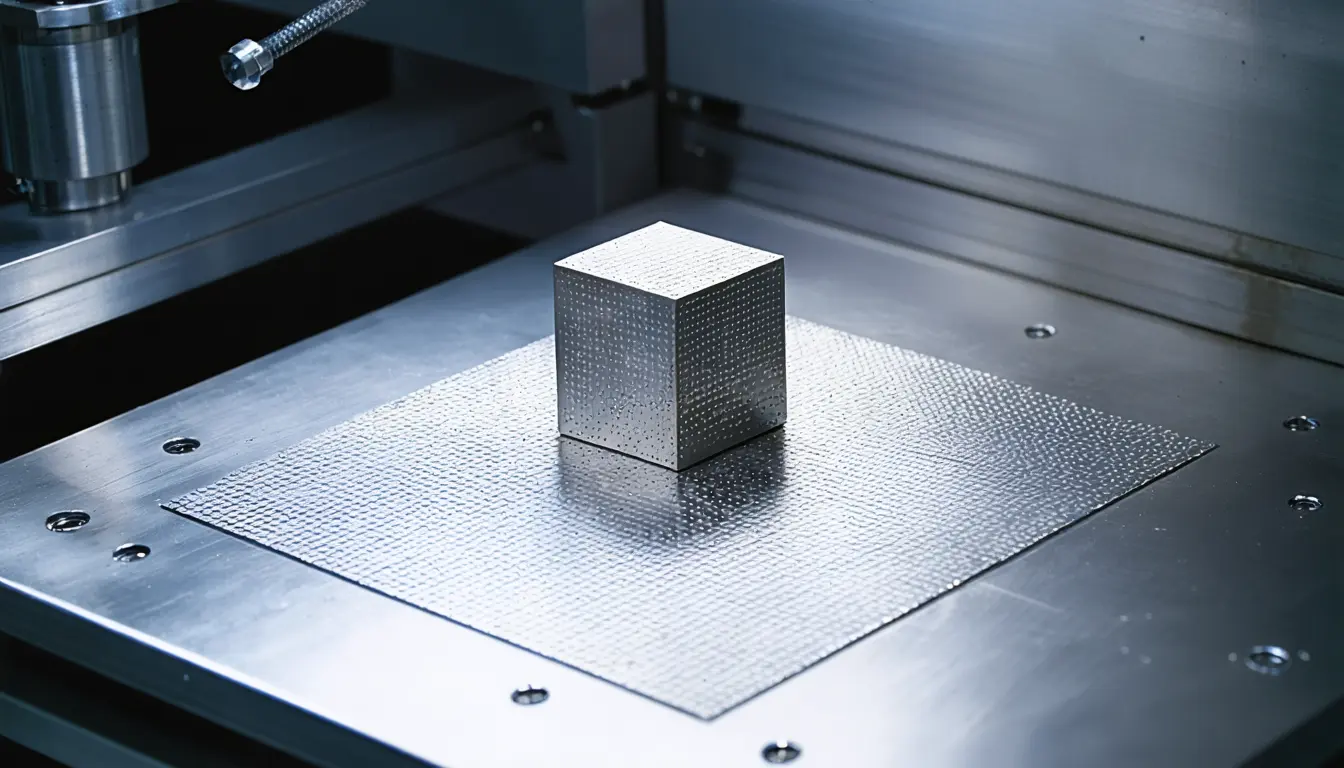
- The team put a randomly patterned tungsten mask in front of the X-ray beam so that each exposure encodes information from several parts of the sample at once.
- They rotated a sample of copper, zirconium and silver while recording the fluorescent X-rays that each element emitted.
- A mathematical algorithm then reconstructed the three-dimensional elemental structures directly from the compressed data.
- According to the researchers, the reconstruction still located microscopic metal wires and particles accurately after more than 97% of the usual measurements were dropped.
Why it matters
- capability At synchrotron facilities, where access is limited, the researchers say a faster scan could cover larger samples, follow chemical processes live and expose delicate materials to less X-ray dose.
- cost Measurement time is saved at the beamline and spent in the reconstruction, so a like-for-like comparison has to count the algorithm's running time as well.
- precedent The team plans to add AI and deep learning to the reconstruction, so later speed and resolution claims will rest on software and will need to name the algorithm behind them.
The two headline figures nearly coincide. A 43-fold speedup means a scan finishes in about 2.3% of the time, a cut of roughly 97.7% [12], and the paper reports the data reduction as more than 97% [1]. That fits if scan time tracks the number of measurements, which is our assumption. The study, "Direct volumetric reconstruction for highly compressive X-ray fluorescence ghost tomography," was published in Optica in 2026 [2].
The design makes every reading a blend of locations, because the mask encodes information from several parts of the sample at once [4]. Recovery then depends on the algorithm that separates them. Adi Ben-Yehuda, a Ph.D. student in Sharon Shwartz's laboratory at Bar-Ilan University's Department of Physics [11], said: "The surprising part was how much information we could recover from so little data." [8] The group's earlier work was two-dimensional X-ray fluorescence ghost imaging, and this extends the idea to a full 3D system [10].
Professor Sharon Shwartz of Bar-Ilan University put the design logic this way: "By designing the X-ray illumination together with the computational reconstruction, we can recover detailed three-dimensional chemical information from a remarkably small number of measurements." [9]
The thing this doesn't tell you is what the 43-fold figure was measured against. The phys.org summary says only conventional approaches [1]. It does not report the number of measurements in either scan, the spatial resolution or the reconstruction time.
In our view the 97% figure is the sturdier of the two. It counts measurements on a defined sample, where the researchers report accurately placing microscopic metal wires and particles [7]. The 43-fold figure depends on the baseline chosen.
What to watch
- Whether the Optica paper reports reconstruction time next to measurement time, and which scan the 43-fold figure was measured against.
- A first reconstruction of a biological or geological sample, and whether the data reduction holds on material denser than metal wires and particles.
- Replication by a group outside Bar-Ilan University and its named collaborators.
Clarity's read
What the record supports and how the coverage leans. The claims behind it follow.
Reality
- Evidence58
- Adoption
- Insufficient
- Hype gap+15
- Incentives
- Insufficient
- Confidence55
Claim ledger
Ranked by verification strength, evidence, and original report placement.
- [1]
Researchers at Bar-Ilan University and collaborating institutions developed an X-ray imaging technique that can map the three-dimensional chemical composition of an object 43 times faster than conventional approaches, while using more than 97% less measurement data.
- [2]
The study, A. Ben-Yehuda et al., 'Direct volumetric reconstruction for highly compressive X-ray fluorescence ghost tomography', was published in Optica (2026).
- [3]
Creating a three-dimensional elemental map with traditional X-ray fluorescence imaging usually requires scanning an object point by point, a process that can take considerable time and generate enormous amounts of data.
- [4]
The researchers placed a randomly patterned tungsten mask in front of the X-ray beam, creating a structured pattern that encodes information from multiple parts of the sample at once.
- [5]
They rotated a sample containing copper, zirconium and silver while measuring the fluorescent X-rays emitted by the different elements.
- [6]
A mathematical algorithm reconstructed the three-dimensional elemental structures directly from the compressed data.
- [7]
The researchers eliminated more than 97% of the measurements normally required while still accurately reconstructing the locations of microscopic metal wires and particles.
- [8]
"The surprising part was how much information we could recover from so little data."
- [9]
"By designing the X-ray illumination together with the computational reconstruction, we can recover detailed three-dimensional chemical information from a remarkably small number of measurements."
- [10]
The technique builds on the team's previous work on two-dimensional X-ray fluorescence ghost imaging, extending the concept into a full 3D imaging system.
- [11]
Adi Ben-Yehuda is a Ph.D. student in professor Sharon Shwartz's laboratory at Bar-Ilan University's Department of Physics.
- [12]
A 43-fold speedup equals a scan taking about 2.3% of the original time, a cut of about 97.7%, close to the reported data reduction of more than 97%.
- [13]
Synchrotron facilities use extremely bright X-ray beams to study materials at microscopic scales, and access to them is limited, making measurement time particularly valuable.
- [14]
Faster imaging could allow researchers to scan larger samples, follow chemical processes as they happen and study delicate materials while reducing their exposure to X-rays.
- [15]
The researchers plan to apply the technique to more complex biological and geological samples and to incorporate artificial intelligence and deep learning to further improve imaging speed and resolution.
Sources
1 independent publisher whose own reporting we read for this story.
- phys.orgPatterned X-rays map 3D chemical structures faster with fewer measurements
1 article · October 13, 2026
Topics and entities
Follow any of these and your For You feed starts watching them — no settings page required.