Science1 publisher3 min readPublished Updated
Myelin repair cells make their own stress hormone, and it seems to set the clock
Max Planck researchers report that oligodendrocyte precursors release CRH near brain injury for about three days. Remove its receptor and you get more precursors but fewer mature cells.
The Scientist · Science desk
Drafted by a language model from the sources cited here and checked against its claim ledger before publication. How we use AISend a correction
What happened
- Oligodendrocyte progenitor cells (OPCs) can mature into oligodendrocytes, which produce the myelin sheath surrounding axons.
- The findings have been published in the journal Cell Reports; the ScienceDaily release is dated August 14, 2026, with the Max Planck Institute of Psychiatry as source.
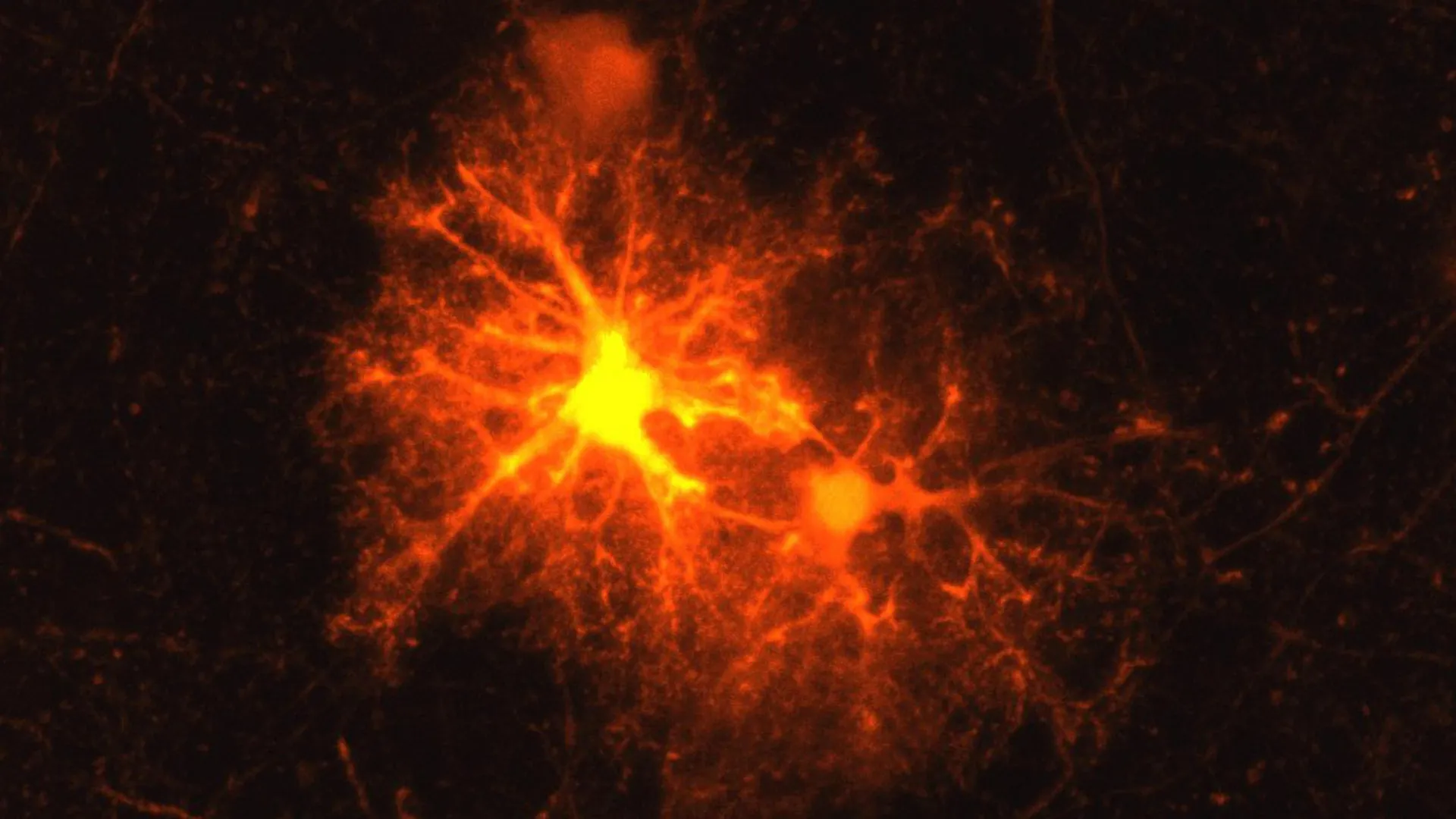
- Near damaged brain tissue, about one third of OPCs activate corticotropin-releasing hormone (CRH), a hormone central to regulating the body's stress response.
- Jan Deussing, a research group leader and neurobiologist, had repeatedly observed a particular group of cells appearing and becoming active around damaged areas in laboratory mice after brain damage such as from an injection, without knowing the cell type.
- Clemens Ries joined the Max Planck Institute of Psychiatry for an internship near the end of his biology degree and took on the question as a master's student; the topic later became his doctoral thesis.
Compiled by The ScientistSomething wrong?How this is made
Why it matters
A group at the Max Planck Institute of Psychiatry reports in Cell Reports that oligodendrocyte progenitor cells, the precursors that build myelin, themselves switch on corticotropin-releasing hormone near damaged brain tissue in mice [1][2][3]. If that holds up, CRH stops being only the opening move of the endocrine stress axis and becomes a local timing signal inside a repair lineage, which is a different kind of drug target than the one demyelinating-disease programs have been arguing about [3][9][10].
The work started as a nuisance observation. Group leader Jan Deussing had repeatedly seen a population of cells turn up and become active around brain damage in laboratory mice, including damage as mundane as an injection, without knowing what the cells were [4]. Clemens Ries, then a master's student on an internship at the institute, tested markers for all known cell types; only the OPC marker responded [5][6]. OPCs mature into oligodendrocytes, which produce the myelin sheath around axons, and that sheath both speeds signal transmission and helps supply the axon with nutrients [1][7]. It breaks down in autoimmune disease such as multiple sclerosis, and physical injury can damage it badly enough that whole neurons die [8].
The numbers are the interesting part. Around wound edges, OPCs proliferate sharply and most go on to mature [9]. About one third of the OPCs near damaged tissue activate CRH [3], which by arithmetic leaves roughly two thirds that do not [2]. The burst is detectable within a few hours of injury and shuts off after roughly three days [10], a window of about 72 hours [1]. Researchers had not previously known that OPCs could produce neuropeptides such as CRH at all [11].
The receptor arm is where the consequence sits. CRH receptor 1 sits on a different population of OPCs, which is what lets those cells respond to the released hormone [12]. Without CRHR1, OPCs multiply faster after injury, but the head start does not convert: fewer mature oligodendrocytes are produced and retained [13]. The authors read this as CRH regulating the timing of maturation, with that timing required to end up with enough mature cells to rebuild the sheath [14]. The operational lesson is blunter. Proliferation and repair moved in opposite directions in the same animals, so counting precursors is not a proxy for remyelination [3].
The institute's summary extends the same system beyond injury, to brain development and to myelin thickness later in life, and suggests it could bear on how early-life stress contributes to psychiatric disorders [15]. That is plausible on the anatomy: CRHR1 is on OPCs even with no injury present, and much of myelination happens after birth and continues into young adulthood [16][17]. It is also the part of the release that is thinnest, and the supplied text breaks off mid-sentence in that section.
Caveats worth holding. This is mouse work from one group, in an injury model whose canonical insult is a needle track, not autoimmune demyelination [4][9]. The release describes no compound tested and no human tissue.
What to watch: whether the mature-oligodendrocyte deficit in CRHR1-null animals reproduces in disease-relevant demyelination rather than mechanical injury; whether the CRH-positive third is a stable subpopulation or a transient state; and whether the roughly three-day window implies that any CRHR1-directed intervention has to be timed rather than continuous [3][10][13][1]. Existing CRHR1 pharmacology was built for the stress axis, so anyone repurposing it will need to show which direction helps, and when.