Science1 publisher2 min readPublished
Cells rush cholesterol into lysosomes to keep them from bursting during mitochondrial cleanup
NTU Singapore researchers report that cells shuttle cholesterol into lysosomes to keep them acidic and intact while digesting damaged mitochondria. The suggested link to Parkinson's and Alzheimer's is still an inference from cell experiments.
The Scientist · Science desk
Drafted by a language model from the sources cited here and checked against its claim ledger before publication. How we use AISend a correction
What happened
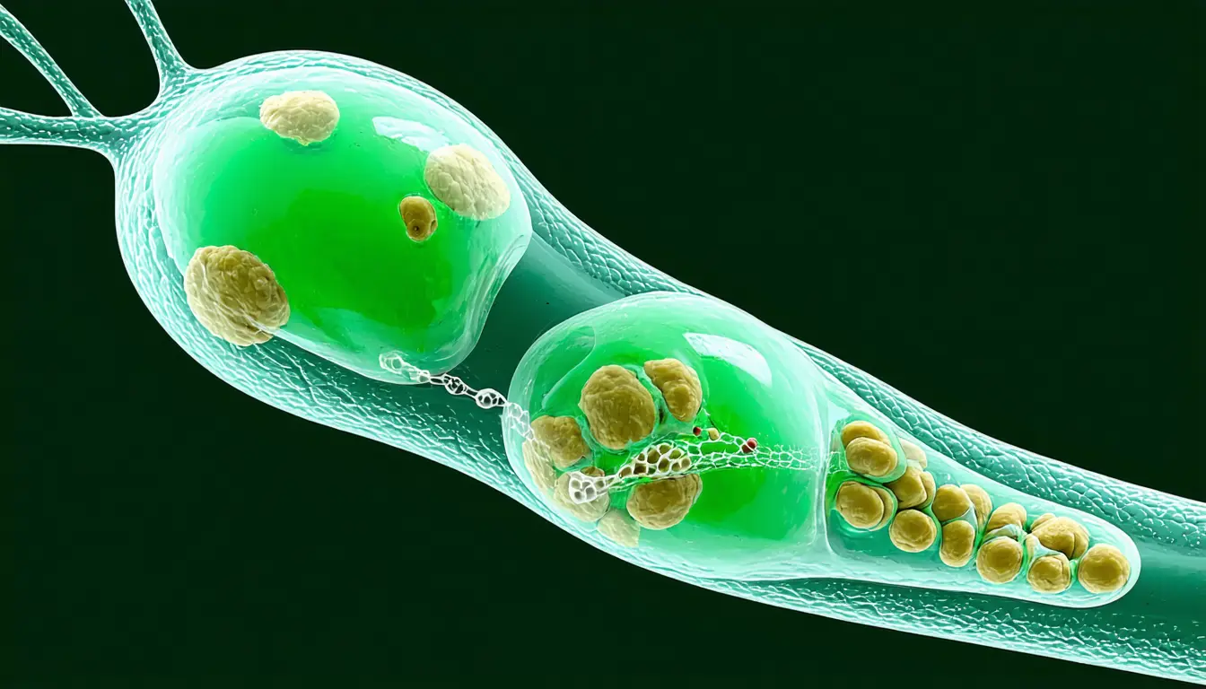
- When damaged mitochondria enter a lysosome, the enzyme PI4KIIalpha marks its surface with the lipid PI4P, which recruits the protein OSBP to carry cholesterol over from the endoplasmic reticulum.
- As cholesterol drains from the endoplasmic reticulum, the cell senses a temporary shortage and turns on a master genetic switch that raises cholesterol production.
- Digesting mitochondrial membranes releases free fatty acids that can become toxic, and cells use a series of enzymes to pack them into lipid droplets as stored energy.
- Blocking cholesterol transport or synthesis left lysosomes less acidic, fragile and unable to clear damaged mitochondria, and the cells formed no lipid droplets.
Compiled by The ScientistSomething wrong?How this is made
Why it matters
- capability The relay gives researchers three named steps, PI4KIIalpha, PI4P and OSBP, that can each be measured in brain tissue to test whether and where the process breaks in disease.
- exposure If the relay works the same way in neurons, those cells would be the most exposed to its failure, since they depend heavily on mitochondrial recycling and cannot easily replace themselves.
- decision Anyone pursuing these steps as drug targets needs a gain-of-function result, because the experiments described show that removing cholesterol harms lysosomes and do not test whether adding more helps.
The study, led from the Lee Kong Chian School of Medicine at NTU Singapore [1], has two parts. First, co-first authors Yang Haoning and Koji Matsuhisa, now an associate professor at Nagasaki University, used live-cell imaging to follow each molecular step as damaged mitochondria arrived in lysosomes [3]. Imaging shows order and location. It can place cholesterol at the lysosome membrane at the right moment, but it cannot show that the lysosome needs it there.
The blocking experiments address that. The team cut off cholesterol at two separate points, its transport and its synthesis, and according to the release both left the lysosomes failing [8]. When two different interventions produce the same failure, a side effect of any single intervention becomes a weaker explanation. In the cells studied, the result supports cause, where the imaging alone showed only timing.
Yasunori Saheki, the corresponding author and an associate professor at LKCMedicine, said, "Lysosomes need to maintain an extremely acidic interior to digest cellular waste effectively. We discovered that as lysosomes take in damaged mitochondria, they reinforce their membranes with cholesterol. This process helps keep the lysosome resilient and ensures that its digestive functions remain fully active." [6]
The release says the findings hold important implications for Parkinson's and Alzheimer's disease [10]. In my view the cell result is persuasive for what it claims, and the disease link is a hypothesis about where to look next. The release does not name the cell types used, report experiments in neurons or disease models, or give effect sizes for the loss of acidity [1]. The paper's own title makes the narrower claim, that cholesterol "maintains the degradative capacity of lysosomes during clearance and recycling of dysfunctional mitochondria" [11].
Yang, a research fellow at LKCMedicine, said, "By mapping how lipid transfer protects lysosomes during mitochondrial damage, we open up potential new therapeutic targets aimed at preserving cellular health during aging." [9]
What to watch
- Whether the PI4KIIalpha-PI4P-OSBP relay operates in neurons, and whether it is weakened in Parkinson's or Alzheimer's models or patient tissue.
- The full Nature Communications paper's methods: which cell types were used and how large the losses of acidity and mitochondrial clearance were.