Science1 publisher3 min readPublished
Synapse PET puts schizophrenia's connection loss on a map, with a left frontal anchor
A Rutgers-led study of 122 people found synaptic loss that follows the brain's chemistry and wiring, not a diffuse smear, and modelling points to a left frontal starting point.
The Scientist · Science desk
Drafted by a language model from the sources cited here and checked against its claim ledger before publication. How we use AISend a correction
What happened
- A study including a Rutgers professor used specialized positron emission tomography (PET) imaging to directly measure synaptic connections in the living human brain; it was published in Molecular Psychiatry.
- The research involved 122 people, including 29 diagnosed with schizophrenia, making it one of the largest synaptic density PET imaging studies conducted so far.
- Participants without a schizophrenia diagnosis numbered 93.
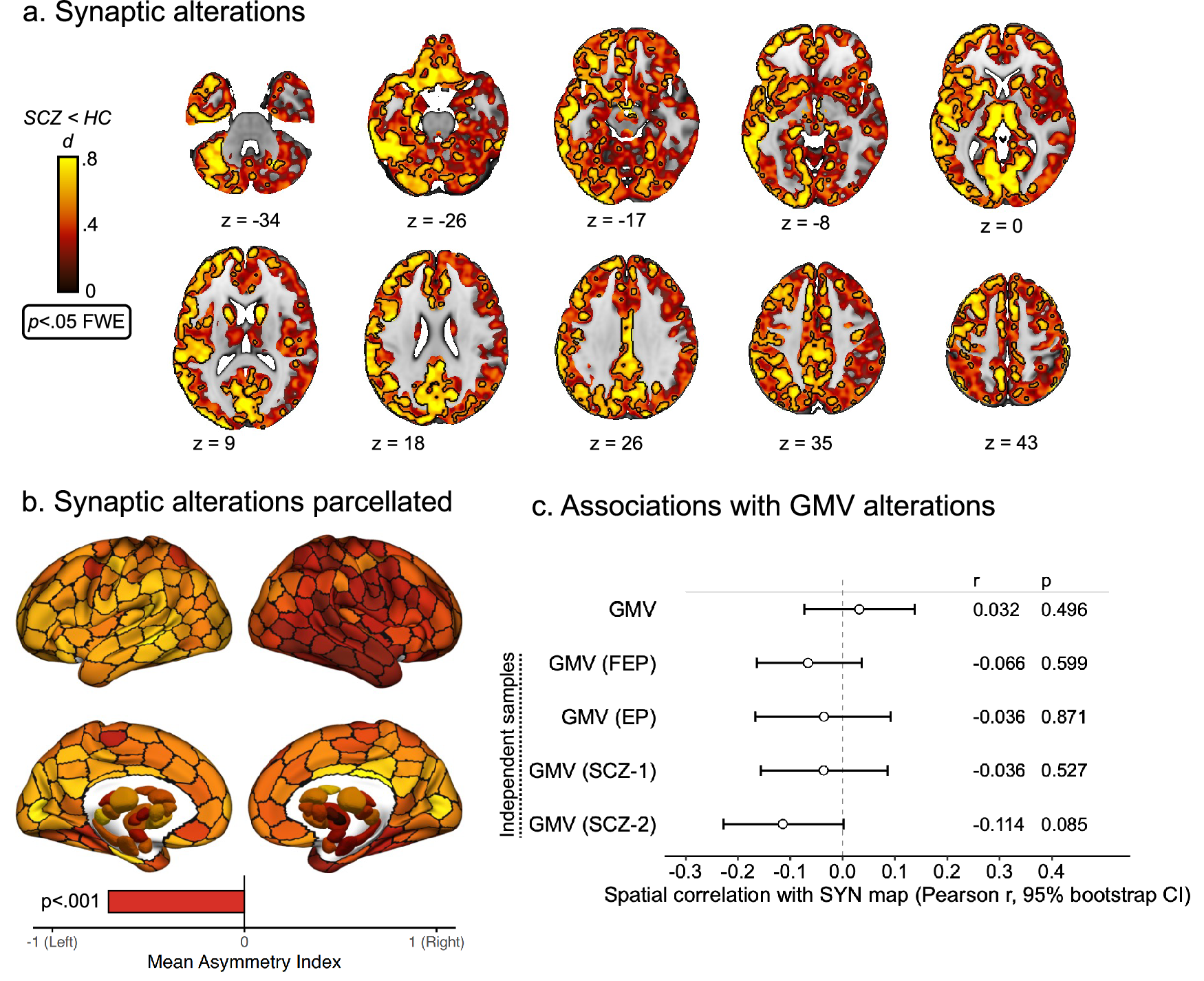
- Compared with healthy participants, people with schizophrenia showed a pronounced and widespread reduction in synaptic connections across several parts of the brain, including frontal and temporal regions and areas involved in memory and emotion.
- The synaptic loss was considerably greater on the left side of the brain than on the right.
Compiled by The ScientistSomething wrong?How this is made
Why it matters
Rutgers-led researchers reported in Molecular Psychiatry that PET imaging of 122 people, 29 of them diagnosed with schizophrenia, found pronounced and widespread reductions in synapses across frontal and temporal regions and areas involved in memory and emotion, with the left hemisphere considerably harder hit than the right [1][2][4][5]. The consequence is not the finding of loss, which was expected, but its geometry: computer simulations based on the brain's structural connections identified an area of the left frontal lobe as a likely starting point from which loss could spread into connected regions [8].
The method is the reason this is new. Conventional MRI cannot specifically measure synapses, which has left the question of where synaptic loss actually occurs in living patients largely unanswered [12]. The team measured those junctions directly with PET, in what the announcement describes as one of the largest synaptic density PET studies conducted so far [1][2]. Scale is relative here: 29 patients is a large clinical sample for this modality and a small one in absolute terms, so the regional and hemispheric contrasts rest on fewer than 30 cases against the remaining 93 participants [3][15].
The most operationally useful result is a dissociation. The synaptic pattern did not match the brain volume changes typically seen on standard MRI, which the authors read as evidence that synaptic loss and volume loss may reflect separate biological processes rather than one process seen through two instruments [6]. Anyone treating structural MRI atrophy as a stand-in for synaptic health should note that.
The spatial organisation also tracks chemistry. Regions with the greatest synaptic losses tended to contain high concentrations of receptors for serotonin, gamma-aminobutyric acid and glutamate, suggesting that a region's molecular makeup shapes how vulnerable it is [7]. "These findings suggest that in schizophrenia, synaptic loss is not random," said first author Sidhant Chopra, now at Orygen and the University of Melbourne, adding that it follows the brain's molecular and connectivity architecture, "which could eventually help identify where and how to intervene" [9][11]. Senior authors were Avram Holmes of Rutgers Robert Wood Johnson Medical School and Rajiv Radhakrishnan of Yale [11].
Two limits are worth holding. The left frontal epicentre is an inference from simulated spread over structural connectivity, not observed propagation in patients [8]. And the release, dated 20 August 2026, does not report participants' medication status, illness duration or symptom severity, all of which bear on whether the pattern reflects the disease, its treatment or its chronicity [14][16].
What to watch is whether the map moves. The researchers say future work will examine how synaptic loss changes over time and how it responds to clinical treatment [13]. If serial scans show loss expanding outward from the same left frontal seed, the process becomes stageable and the epicentre becomes a target; Holmes framed the mapping as a step toward identifying where to intervene, including with emerging therapies meant to prevent and regrow synapses [10]. If different patients turn out to have different epicentres, the useful unit stops being schizophrenia and starts being the individual scan.