Science1 publisher3 min readPublished
Göttingen team separates eight proteins in one cell by how long each dye keeps glowing
Fluorescence lifetime gives a second axis for separating labels. A Göttingen-led team used it to read eight proteins from a single stained cell on an ordinary confocal with lifetime detection.
The Scientist · Science desk
What happened
- An international team led by University Medical Center Goettingen reported in ACS Nano that it visualised eight different proteins in one cell simultaneously, in a single staining step, on standard microscopes.
- Conventional fluorescence microscopes detect only three or four colours at a time, so a sample has until now yielded only three or four distinguishable protein labels.
- The group measured 26 dyes across the four pocket variants and compiled a catalog for picking workable combinations; it calls the method NanoFLex.
Compiled by The ScientistSomething wrong?How this is made
Why it matters
- cost Laboratories that already run a confocal with fluorescence lifetime detection can add labels without buying an instrument, and according to Tsukanov no specialized platform is needed; those without lifetime detection now have a detector purchase to justify.
- capability Antibody stocks a laboratory has already validated can be given a lifetime code by the nanobody, so adding targets no longer waits on newly conjugated antibodies.
- constraint The limit on multiplexing moves off the detector's colour channels and onto dye chemistry and computational unmixing, and the authors tie any further increase to both.
- precedent Pathology gets a credible route to many markers from a single scarce section, because tissue autofluorescence can be separated by lifetime; the authors put this forward as an expectation for clinical samples.
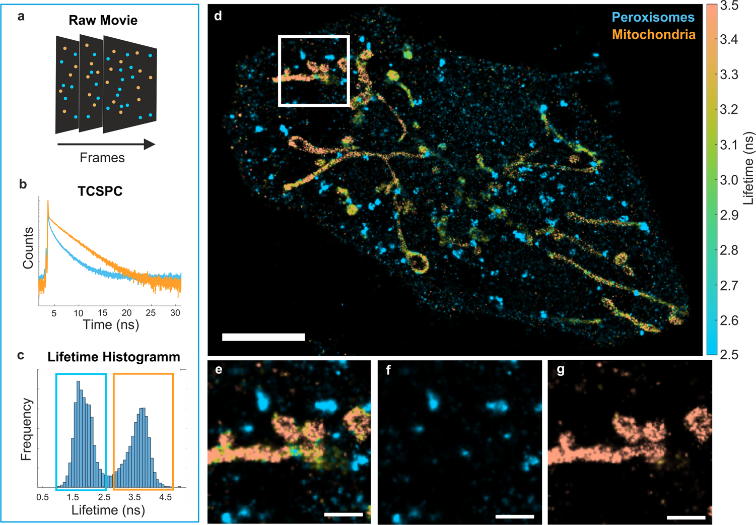
The lifetime depends on a dye's immediate chemical surroundings, and the surroundings are what the Göttingen group engineered [9]. Each dye molecule sits in a protein pocket that exists in four genetically modified variants, and each variant hands the same dye a different lifetime [9]. A single red dye in four pockets gives four labels. A colour detector cannot separate them; a timing detector can [10].
"Two labels can look identical in color and still be cleanly separated, because one fluoresces measurably longer than the other. It is a bit like telling two instruments apart not by the pitch of the note, but by how quickly the sound fades away," said Felipe Opazo, the UMG group leader who conceived and led the study [7][5].
Four lifetimes per colour and eight reported targets means at least two colour channels were in play, since 4 x 2 = 8 [21]. phys.org did not report a misassignment rate or the margin separating the four lifetimes [23]. Those numbers decide whether eight targets hold up in a thick, dim sample, and they are the first thing I would look for in the paper.
A lifetime code is usable only with a lookup table, so the team measured 26 dyes against all four pocket variants and compiled a catalog of combinations to select from [14]. 104 dye-pocket measurements in all [20]. They called the method NanoFLex, for nanobody-guided fluorescence lifetime multiplexing [15].
Delivery is the practical part. The pockets are coupled to nanobodies, alpaca-derived fragments about a tenth the size of a conventional antibody [11]. A conventional antibody finds the protein, the nanobody finds the antibody, and the lifetime code travels with it, so an antibody a lab already uses can be given one [12]. Because the two are preassembled before staining, all targets go on in a single step [13]. In my view that step matters more than the number eight: it is what will decide whether anyone outside Göttingen runs it.
"It was important to us that the method can be readily implemented on conventional confocal microscopes equipped with fluorescence lifetime detection. It requires no specialized microscopy platform," said Roman Tsukanov of the Third Institute of Physics at the University of Göttingen, a senior author of the study [8][5]. His sentence contains a condition: the confocal needs lifetime detection [8].
Human tissue glows on its own, and that background interferes with many staining methods, but its lifetime is easy to tell apart from a real signal [16]. The authors write that the approach could prove particularly useful for clinical samples, where reading many markers from one scarce section would matter [17]. The work raises that as a prospect and does not demonstrate it [17]. They expect the count of simultaneously detectable proteins to rise as new dyes and better computational analysis become available [18].
What to watch
- Look for misassignment rates and the lifetime separation margins for the eight-target images in the ACS Nano paper.
- Whether the four-variant pocket set grows, since the authors tie higher multiplexing to new dyes and better computational analysis.
- Whether the autofluorescence separation claimed for human tissue holds in an actual biopsy section and not only in cultured cells.