ScienceNot yet confirmed elsewhere1 publisher2 min readPublished
Brown's million-electrode chip follows migrating cancer cells by the capacitance of their grip
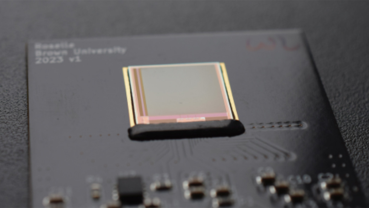
Brown University engineers tracked breast cancer cells on a 1-million-electrode chip by reading capacitance changes, with no dye or optics. Dropping the fluorescent label could let labs studying metastasis watch cells without the dye exposure a co-author says can damage DNA.
The Scientist · Science desk
What happened
- Cells gripping the chip to move change the electrical properties at its surface, and the array follows each cell through those small shifts in capacitance.
- In laboratory tests the chip also captured cell division events as they happened.
- With multicellular spheroids, it recorded leader cells at the clusters' outer edges as they began migrating outward.
- It imaged honeycomb tissues of hundreds of thousands of cells, resolving the voids and boundaries left by complex changes in shape.
- The work is published in the journal Lab on a Chip.
Why it matters
- constraint Experiments that need to see more than where a cell touches the surface still need optics, because contact is the only thing this sensor reads.
- capability Tissues big enough to need several stitched microscope frames fit in one sensor field, so collective-migration studies drop an image-alignment step.
- decision A lab weighing the chip against a microscope it already owns is working from the team's statement that it could be mass-produced fairly inexpensively, with no unit price yet attached.
Jacob Rosenstein, an associate professor of engineering who co-led the study, compares the chip to a fingerprint scanner. "There are differences in electrical capacitance between the ridges in a fingerprint and the spaces in between, and that's what some scanners read," he said [16]. "It's a similar concept here. We're detecting the differences in electrical capacitance between where cells are touching the surface and where they are not, as they move over time." [10] The paper's title describes the device as a megapixel CMOS capacitance sensor [15].
Two published figures, the chip's size and its electrode count, set an upper bound on each pixel. A square chip about 1.3 centimetres on a side has roughly 1.69 square centimetres of surface [8]. Shared among a million electrodes [7], that leaves at most about 169 square micrometres apiece, a square roughly 13 micrometres across [18]. An array cannot be larger than the chip that carries it, so the real spacing is that size or finer.
Hyuntae Jeong, a postdoctoral researcher in engineering at Brown who shares first authorship of the paper, framed the problem as one of labels. "Cells often have to be labeled with fluorescent dyes in order to see them, but dyes can cause DNA damage, which could affect the way they move or even cause cell death," he said [2]. His fellow co-first author, Pushkaraj Joshi, stated the benefit with a hedge: "By using capacitance rather than light, our sensor lets us observe cell migration without labeling, which may decrease unwanted artifacts." [3]
The thing this doesn't tell you is how large that decrease is. Phys.org's write-up of the study does not describe a side-by-side run of dyed and unlabelled cells comparing how fast they move or how often they divide. As reported, the gain for the cells rests on Jeong's account of what dyes can do [2].
The nearer gain, I think, is in the analysis. "Moving cells are very challenging to track since they change shape and move rapidly, so computational analysis can be quite error-prone," Jeong said [5]. Ian Y. Wong, an associate professor of engineering who co-led the project, described the chip's output differently. "The cells show up beautifully in the capacitance image and can be tracked automatically," he said. "These videos are so much easier to analyze than what we typically get from fluorescence microscopy, where we always have to fine-tune the image processing." [4]
The hardware is also small. "One of the reasons we think this is interesting is that it has no lenses or light sources or optics at all, so it's very small and lightweight," Rosenstein said [20].
What to watch
- A head-to-head run of one cell line, dyed under a microscope and unlabelled on the chip, comparing migration speed and division rates.
- Tests with immune cells or wound-healing models, which the Brown team names as target processes but which the reported experiments did not include.
Clarity's read
What the record supports and how the coverage leans. The claims behind it follow.
Reality
- Evidence50
- Adoption
- Insufficient
- Hype gap+20
- Incentives40
- Confidence55
Claim ledger
Ranked by verification strength, evidence, and original report placement.
- [1]
The system tracks cell movement through electrical capacitance, presented as an alternative to optical microscopes and fluorescence imaging, which can damage cells and potentially disrupt their behaviour.
- [2]
"Cells often have to be labeled with fluorescent dyes in order to see them, but dyes can cause DNA damage, which could affect the way they move or even cause cell death."
ReportedSupportedSource: Hyuntae Jeong, postdoctoral research associate, Brown School of Engineering, co-first author2 sources— create a free account to open themView cited source - [3]
"By using capacitance rather than light, our sensor lets us observe cell migration without labeling, which may decrease unwanted artifacts."
ReportedSupportedSource: Pushkaraj Joshi, co-first author, now a senior research associate in Brown's Department of Physics2 sources— create a free account to open themView cited source - [4]
"The cells show up beautifully in the capacitance image and can be tracked automatically. These videos are so much easier to analyze than what we typically get from fluorescence microscopy, where we always have to fine-tune the image processing."
ReportedSupportedSource: Ian Y. Wong, associate professor of engineering, co-led the project2 sources— create a free account to open themView cited source - [5]
"Moving cells are very challenging to track since they change shape and move rapidly, so computational analysis can be quite error-prone."
ReportedSupportedSource: Hyuntae Jeong2 sources— create a free account to open themView cited source - [6]
Engineers from Brown University developed a technique for imaging cell migration dynamics, described in a study published in the journal Lab on a Chip.
- [7]
The technique uses an array of 1 million microscale electrodes on which cells can be placed in a liquid nutrient solution.
- [8]
The microelectrodes are arranged on a square microchip with sides about a half inch (1.3 centimeters) long.
- [9]
When cells placed on the chip grip the surface to move, the contact affects the electrical properties near the chip surface; by measuring tiny changes in capacitance the device tracks each cell as it moves across the array.
- [10]
"It's a similar concept here. We're detecting the differences in electrical capacitance between where cells are touching the surface and where they are not, as they move over time."
ReportedSupportedSource: Jacob Rosenstein, associate professor of engineering, co-led the studyView cited source - [11]
In laboratory tests the technique tracked the motion of human breast cancer cells and captured cell division events as they occurred.
- [12]
The chip revealed the dynamics of multicellular spheroids, capturing the motion of leader cells at the outer edges of clusters as they began migrating outward.
- [13]
The researchers imaged honeycomb tissues composed of hundreds of thousands of cells, showing the technique could resolve voids and boundaries due to complex changes in shape.
- [14]
The relatively large sensor area enables imaging of large tissues without stitching together multiple images, which is often necessary with optical microscopy.
- [15]
The paper is titled 'Label-Free All-Electrical Tracking of Individual and Collective Cell Migration on a Megapixel CMOS Capacitance Sensor' (Jeong et al, Lab on a Chip, 2026).
- [16]
"There are differences in electrical capacitance between the ridges in a fingerprint and the spaces in between, and that's what some scanners read."
- [17]
The Brown team describes cell migration dynamics as important to understanding cancer metastasis, wound healing and immune response.
- [18]
Each of the million electrodes occupies at most about 169 square micrometres of chip area, equivalent to a square roughly 13 micrometres across; this is an upper bound because the array cannot exceed the chip.
- [19]
The researchers say the device could be mass-produced fairly inexpensively.
ReportedInsufficientSource: Brown researchers, as reported by phys.org2 sources— create a free account to open themView cited source - [20]
"One of the reasons we think this is interesting is that it has no lenses or light sources or optics at all, so it's very small and lightweight."
ReportedInsufficientSource: Jacob Rosenstein2 sources— create a free account to open themView cited source
Sources
1 independent publisher whose own reporting we read for this story.
- phys.orgAll-electrical imaging offers innovative alternative for tracking cell movement
1 article · October 8, 2026
Topics and entities
Follow any of these and your For You feed starts watching them — no settings page required.