Science1 publisher2 min readPublished
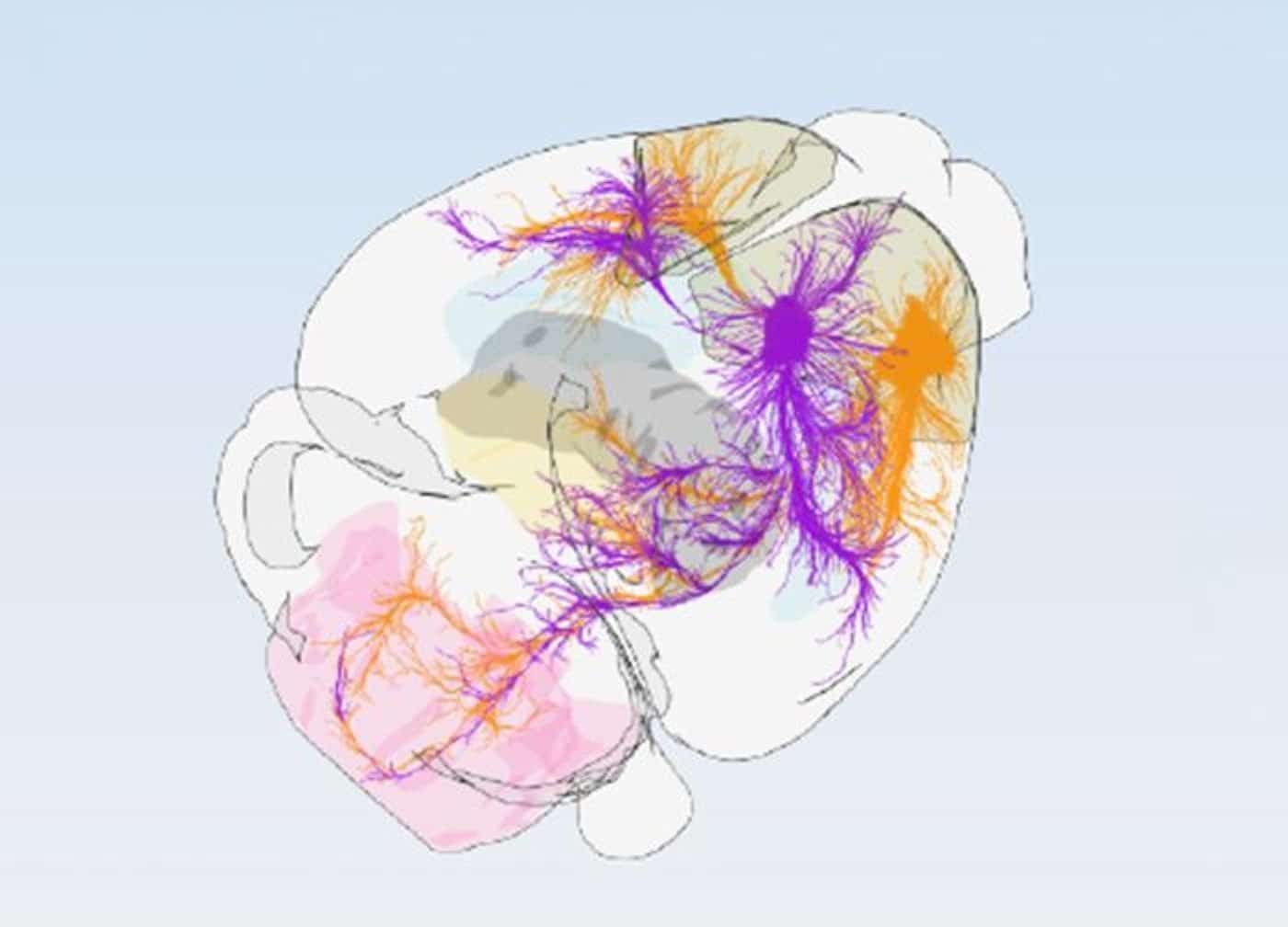
Clustering 547 tracing experiments splits the mouse motor cortex into 16 modules
A Basel, FMI and Allen Institute team grouped cortical sites by where their axons end, and found primary and secondary motor cortex both reaching the brainstem and spinal cord directly. The 16-module layout is now in the open BrainGlobe atlas.
The Scientist · Science desk
What happened
- Researchers clustered 547 projection-tracing datasets from the Allen Institute's Mouse Brain Atlas by where each cortical site sends its axons, and sorted the mouse motor cortex into 16 modules.
- The modules sit in three rows on two axes: anterior sites handle planning and decision-making while posterior ones couple to sensory feedback, and the medial-to-lateral axis runs from trunk and limbs to jaw, mouth and face.
- Primary and secondary motor regions were found projecting in tandem straight into the brainstem and spinal cord, against the classical assumption that M2 relays commands down through M1.
- Single-neuron morphology reconstruction and spatial cell-type profiling were run as independent checks, and both returned the same 16-subregion layout as the tracing analysis.
- The University of Basel, Friedrich Miescher Institute and Allen Institute team has put the 16-subregion blueprint into the open-source BrainGlobe atlas.
Compiled by The ScientistSomething wrong?How this is made
Why it matters
- capability A shared 16-module frame lets two labs reporting different neuronal vulnerability in ALS models check whether they were sampling the same tissue at all.
- constraint Because the modules were cut by projection target, the map places a site precisely without telling anyone what that site computes; physiology still has to be recorded module by module.
- decision Groups that log sites as M1 or M2 now have to decide whether to re-register old coordinates before pooling data with anyone using the finer parcellation.
- contradiction The subregions are described as functionally distinct, yet all three converging methods are anatomical or molecular, so the functional claim is an inference from wiring.
The unit of evidence here is an injection. Each of the 547 datasets is a tracer placed at one set of cortical coordinates in a mouse, with the labelled axons then catalogued across sensory, motor and cognitive targets; sites whose axons land in the same places were grouped together [2]. Sixteen modules out of 547 experiments averages about 34 experiments per module [12].
The hierarchy finding carries the most weight. Classical accounts put M2 above M1, relaying commands downward; the tracing shows both areas sending axons straight into the brainstem and spinal cord [4]. Axon terminals are what that tracing counts. It does not time the two areas during a movement, and none of the three converging methods records activity: axonal tracing, single-neuron morphology reconstruction and spatial cell-type profiling are anatomical or molecular measurements [5][14].
"Understanding the precise wiring of the brain is essential for developing effective treatments for brain diseases," said Hongkui Zeng, Executive Vice President and Director of Brain Science at the Allen Institute and a senior author of the study [8][9].
The framework is offered as standardised anatomy for studying selective neuronal vulnerability in ALS and frontotemporal dementia [10]. For that work, the addresses change. Standard reference atlases carried two compartments, M1 and M2, a division the authors say failed to account for the functional specialisation, cell populations and connection topographies they see [11]; sixteen modules is eight times that [13]. So a methods section reporting "M1" is naming a region the new map divides into several modules with different downstream targets.
The release does not report any reanalysis of earlier recordings or lesions under the new parcellation. On this evidence, older two-zone studies used a label that pools tissue the tracing separates. That does not make them wrong. Whether two labs disagreeing about vulnerability in M1 were sampling the same modules is now a checkable question, and checking it means going back to their coordinates.
Modules were defined by shared projection targets [2]. Each module is therefore a prediction about function. And all 547 tracing datasets came from mice.
What to watch
- Whether functional recordings inside single modules find the distinct tuning that clustering by projection target predicts.
- Whether ALS and frontotemporal dementia papers start reporting recording and lesion sites by module and BrainGlobe coordinates instead of M1 or M2.
- Whether the same dual-axis layout appears when the method is applied to tracing data from other species.