Science1 publisher2 min readPublished
Where a locus coeruleus neuron sits decides where its norepinephrine goes
In a Nature study from the Allen Institute, the mouse locus coeruleus sorts its output by position, with cells high in the nucleus serving cortex and learning and cells low in it serving the brainstem and spinal cord.
The Scientist · Science desk
What happened
- Allen Institute researchers report in Nature that the mouse locus coeruleus sends tailored norepinephrine signals to specific destinations, using brain imaging, electrophysiology, single-cell genetics and behavioural tasks.
- Cells in the upper, dorsal part of the nucleus project into the forebrain and cortex, and they fired when mice revised a decision after negative feedback.
- Cells in the lower, ventral part project down to the brainstem and spinal cord, and their firing spiked immediately before an animal chose to ignore a cue offering a possible reward.
Compiled by The ScientistSomething wrong?How this is made
Why it matters
- constraint A single number for norepinephrine level cannot say which tier of the nucleus a drug moved, so the readout most norepinephrine pharmacology relies on is blind to the division this map draws.
- decision Programs aimed at norepinephrine now have a choice of target unit, since the Allen Institute offers the map as a blueprint for pathway-specific therapy instead of whole-brain flooding.
- capability Because locus coeruleus cells die early in Alzheimer's, whether the dorsal tier thins before the ventral one becomes a question tissue studies can now ask with a coordinate to test against.
Position inside the nucleus predicts two things at once, where a cell sends its axon and which genes it turns on. "What emerged was a clear map: neurons in the dorsal LC that send signals upward to the cortex are involved in learning, whereas neurons in the ventral region projecting downward to the brainstem and spinal cord govern whether animals engage with their environment at all," said Karel Svoboda, director of Neural Dynamics at the Allen Institute and a co-author. "We also showed that these anatomical differences are mirrored by distinct gene expression patterns." [6][14]
The separation therefore shows up three times over, in the wiring, in the firing during a task, and in the transcriptome [1]. For decades the working model held that the locus coeruleus released a generalized surge of norepinephrine across the whole brain whenever an animal met stress or novelty [2]. "The findings suggest the brain's norepinephrine system is far more like a targeted postal network than a foghorn," Svoboda said [5].
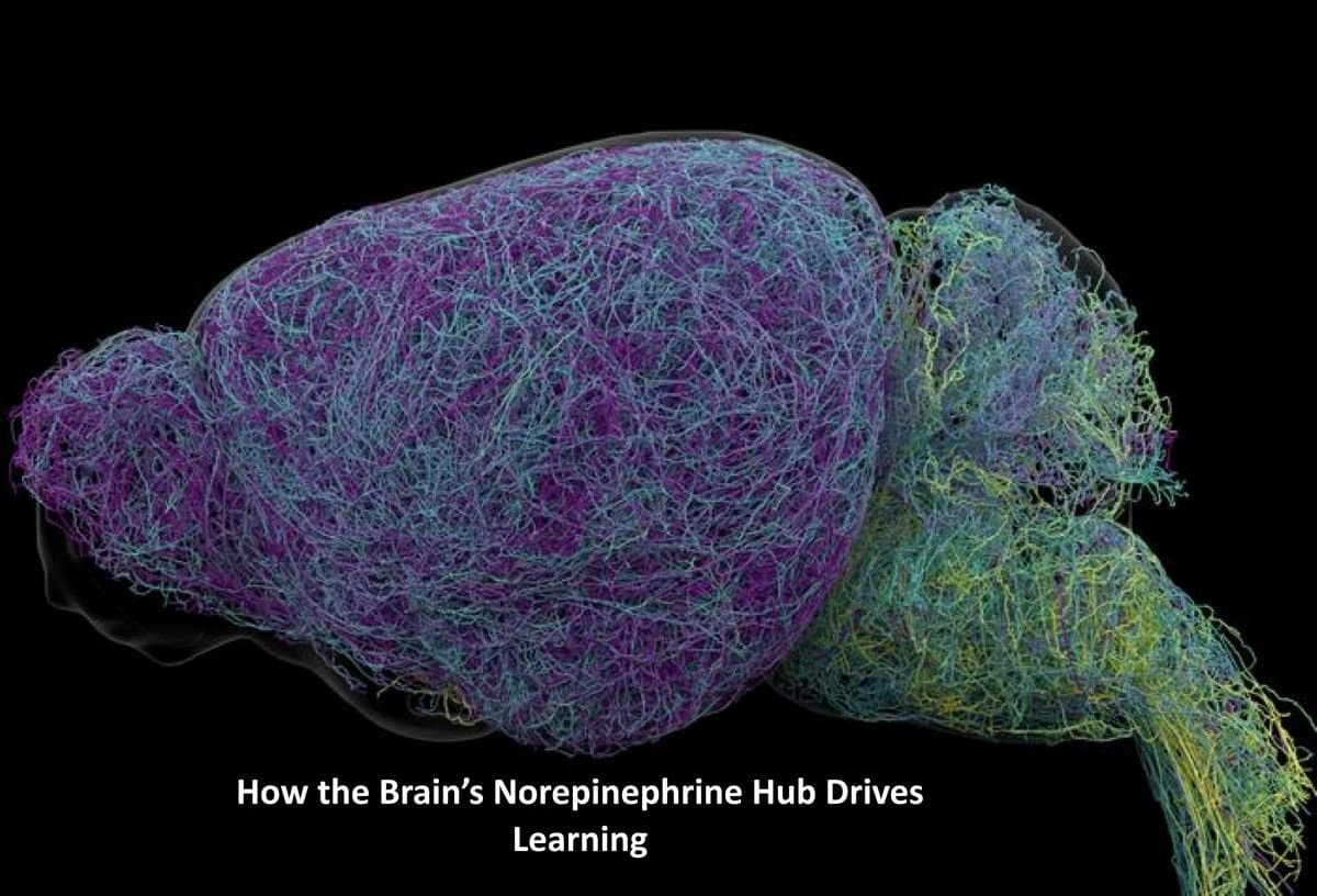
Start with the morphology. Complete reconstructions of individual projections came out of whole-brain imaging across nearly 35,000 neurons, with roughly 400,000 cells profiled genetically alongside them [7]. Axons in the locus coeruleus average about 35 centimetres [8]. One measured 70.32 centimetres, twice the average, and the team reports it as the longest single neuron documented in a mouse [9][13]. The paper does not give the sample size for that average; the figure released with the study superimposes about 100 of them, coloured by where in the nucleus they sat [10][15].
Locus coeruleus cells degenerate early in Alzheimer's disease, and norepinephrine pathways are primary targets for ADHD, depression and anxiety drugs. The Allen Institute presents this circuit map as a blueprint for therapies aimed at specific pathways instead of flooding the entire brain [12]. The nucleus is a tiny fraction of the brain's cells and the main source of a transmitter that governs attention, stress, arousal, heart rate and learning [11]. If the two tiers do different jobs, a readout that reports one norepinephrine number cannot identify which tier a compound moved [3][4].
On the ventral side the evidence is timing. Firing rose immediately before mice chose to disengage from or ignore cues offering a possible reward [4]. The dorsal cells get stronger language: the account describes them as actively driving learning and behavioural adaptation when mice updated decisions after negative feedback [3]. All of it was measured in mice.
What to watch
- Whether silencing the dorsal or ventral tier alone breaks learning or engagement selectively. A selective break would make the ventral timing result a causal one.
- Whether the dorsal and ventral gene expression differences yield markers usable as drivers for pathway-specific compounds.
- Whether human post-mortem locus coeruleus tissue in Alzheimer's shows the same dorsal-ventral division, and which tier thins first.