Science1 publisher3 min readPublished
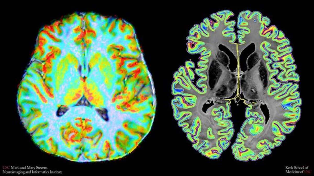
USC pairs 7T perfusion with a histology atlas to score blood supply layer by layer
Researchers at USC scanned 30 adults at 7 Tesla and matched laminar blood flow against cell-body staining from the BigBrain atlas, producing an index that tracks mitochondrial respiratory capacity across 360 cortical regions.
The Scientist · Science desk
What happened
- Researchers at USC's Stevens INI built a noninvasive index, CCSI, that compares layer-by-layer cortical blood perfusion with cell packing across 360 cortical parcels, in a paper published in Nature Communications.
- They scanned 30 healthy adults using arterial spin labeling on a 7 Tesla scanner at one cubic millimeter resolution, and 14 of them returned for a second session to check test-retest reliability.
- Across most of the cortex, perfusion tracked cell density, with more densely packed layers receiving proportionally greater blood flow.
Compiled by The ScientistSomething wrong?How this is made
Why it matters
- capability A group already running arterial spin labeling at 7 Tesla can compute a layer-resolved supply-and-demand number from scans it collects anyway, with no injected tracer and no extra contrast agent.
- contradiction USC's summary states the cognitive finding at two strengths, and only the narrower one, better prediction of fMRI activity in association cortex, matches what was measured.
- decision Any lab that wants to use CCSI as a per-person marker has to fund its own reliability work, because 14 repeat scans at a single site is where the published evidence stops.
Arterial spin labeling magnetically tags water molecules in arterial blood and uses them as an endogenous tracer, so the perfusion maps need no injected contrast [4]. At 7 Tesla the USC team held that measurement to one cubic millimeter isotropic voxels [4], fine enough to sample perfusion from the outer cortical surface inward instead of collapsing it into one value per region [6]. Averaging across the full cortical thickness is what conventional vascular imaging has done, and it hides how microcirculation meets the needs of particular layers [11]. "Conventional brain imaging often averages information across the full thickness of the cortex, but the cortex is not a uniform sheet," said Chenyang Zhao, co-first author of the study [14].
The cellular half of the index is not measured in the volunteer. Cell-body staining intensity comes from BigBrain, a 3D histological reconstruction of cellular packing used here as a reference atlas [16]. Perfusion comes from each participant's own scan [5]. The cell-density term is therefore the same for everyone in the sample, and whatever spread CCSI shows between people comes from the blood side [20].
Cortical tissue has virtually no capacity to store energy and depends on continuous delivery of oxygen and glucose [21]. Higher alignment between flow and cell density went with greater mitochondrial respiratory capacity, and not with mitochondrial volume or with raw bulk blood flow [7]. The metabolic and genetic references used for that comparison are atlases [17], and CCSI itself is defined over 360 cortical parcels [3], so the finding describes where in the cortex supply and packing line up. Whether the alignment produces the respiratory capacity, follows from it, or shares an upstream cause is not settled by a spatial correlation.
USC's summary carries the cognitive result at two strengths. One line says CCSI significantly improved models predicting higher-order cognitive brain function [9]. The key-facts list says adding CCSI to structure-function coupling models improved prediction of neural activity in association areas responsible for memory, reasoning and attention [8]. The second is the measured one. Predicting fMRI activity in association cortex is a different task from predicting how well a person remembers or reasons, and the summary gives no cognitive test scores and no size for the improvement [22].
For a group that already runs ASL at 7 Tesla, the draw is a layer-resolved supply-and-demand number from a sequence it collects anyway [4]. Repeatability rests on the 14 of 30 volunteers who came back for a second session [5], 47 percent of the sample [18]. "Our new measure gives us a way to study how well that energy supply is positioned to meet cellular demands in different parts of the cortex," said Fanhua Guo, co-first author and a researcher at Stevens INI [13].
What to watch
- Whether an independent 7T site reproduces CCSI's test-retest reliability, and whether any version of it survives at 3 Tesla.
- A study that pairs CCSI with actual cognitive testing rather than with fMRI activity in association cortex.
- Substituting per-subject cellular or metabolic measurements for the group atlases, in aging or small-vessel disease cohorts.