Science1 publisher2 min readPublished
EPFL's lipid atlas splits the mouse brain into 539 chemical territories
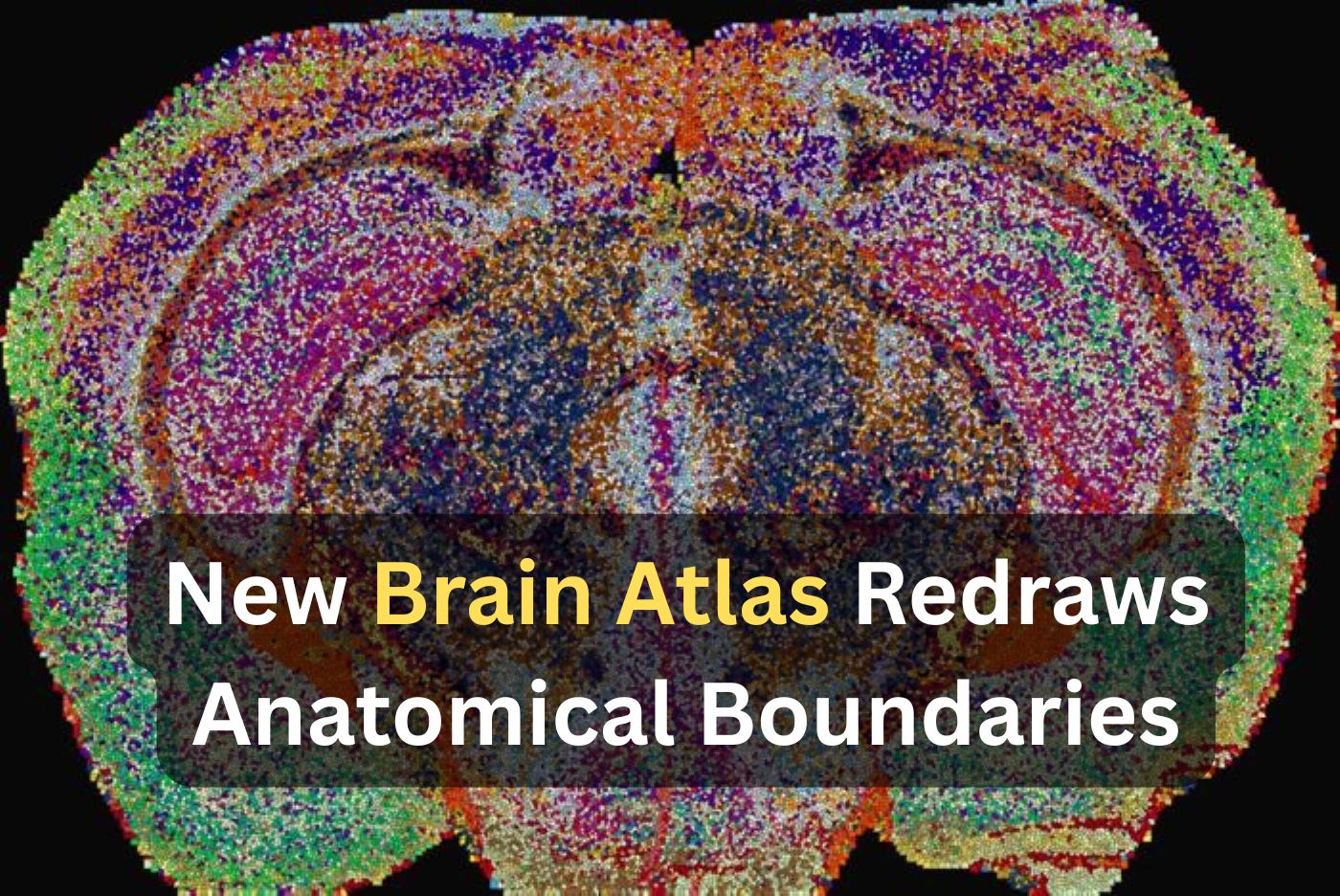
Researchers at EPFL imaged 172 lipids in 109 slices from 11 mice and sorted the readings into 539 zones. The zones follow known anatomy and also subdivide white matter that gene-expression maps treat as uniform.
The Scientist · Science desk
What happened
- An EPFL team's Lipid Brain Atlas, published in Nature, divides the whole mouse brain into 539 territories it calls lipizones, each defined only by its own combination of lipids.
- The map was built with mass spectrometry imaging, which lasers thin brain slices point by point, covering 172 lipids across 109 slices taken from 11 mice.
- Turning roughly seven million individual readings into one 3D model of the brain required machine-learning tools the team wrote for the scale of this dataset.
- The signatures were distinctive enough that the researchers could say which region a piece of tissue came from by analysing its lipid composition alone.
- White matter, usually treated as chemically uniform, came out as a patchwork of distinct zones, with myelin-making cells differing biochemically by location.
Compiled by The ScientistSomething wrong?How this is made
Why it matters
- capability A lipid readout can now place mouse tissue anatomically without genes, proteins or stains. That gives a lab a second and independent way to check whether a manipulation has moved a region off its normal chemistry.
- constraint Eleven mouse brains cannot carry a claim about human disease, so any work linking these zones to depression or Alzheimer's has to re-derive the map in human tissue first.
- decision Groups studying myelin now have to decide whether "white matter" is still an acceptable unit of analysis, because where the sampling needle lands may determine which chemical zone the sample represents.
- precedent Lipids can tag cell bodies and their distant terminal fields alike. That gives atlas projects that have relied on transcriptomes a reason to add a lipid channel, to capture relationships gene expression does not encode.
Five hundred and thirty-nine territories came out of 172 measured lipid species, about three zones for every lipid on the list [14]. Identity comes from the combination of the measurements taken at each point [2]. No single molecule labels a lipizone. EPFL did not say how the team settled on 539 as the cluster count, and I would expect a second group to test that number before anything else.
The sampling is uneven in a way the totals hide. Eleven mice yielded 109 slices, roughly ten per animal [13], and the seven million readings work out to around 64,000 per slice [15]. Within a slice the map is dense. Along the front-to-back axis it is built from a much smaller number of planes, pooled across all 11 animals [3][4].
The region-identification result is a classification result: chemistry predicts location well enough to place a piece of tissue [5]. Whether lipids draw the boundaries is a separate question. Membrane composition could equally follow from which cells happen to sit where, and mass spectrometry imaging of fixed slices cannot separate the two.
Some lipizones do something gene-expression maps structurally cannot. They give a cluster of nerve cell bodies and the far-off terminal areas its fibers reach the same chemical label, a relationship that maps built on cell bodies usually miss [6]. "In effect, lipids work like postal codes, giving distant but related parts of the brain the same chemical address," said Giovanni D'Angelo, the EPFL professor who led the work with Luca Fusar Bassini and Gioele La Manno [8][1].
The motivation D'Angelo gives is human. "So scientists have had only a blurry picture of how lipids differ from one part of the brain to another," he said [9]. "That gap matters, because changes in brain lipids are increasingly linked to conditions from depression to Alzheimer's disease" [10]. The atlas itself is 11 mouse brains [3], and whether human white matter carries the same subdivisions is still open. The white-matter patchwork is the finding I would want replicated in human tissue first, because the uniformity it disagrees with is an assumption a great deal of myelin work is built on [7].
What to watch
- Whether the lipizone boundaries, and especially the white-matter subdivisions, reproduce in human post-mortem tissue at comparable resolution.
- Whether the lipid zones inside white matter line up with oligodendrocyte transcriptomic subtypes when both are measured in the same brain.
- Whether the machine-learning tools and the 7 million readings are released in a form other labs can register their own slices against.