Science1 publisher3 min readPublished
Synchrotron scans catch fungal hyphae inside intact soil around tomato roots
Henri Braunmiller's Munich team imaged fungal hyphae inside intact soil using synchrotron CT, in minutes where a lab CT can take hours. The 3D views place hyphae among roots and sand grains in small potted samples.
The Scientist · Science desk
What happened
- The team grew tomato plants in loam and in sandy soil seeded with fungal spores, hyphae and colonized root fragments.
- Perforated soil cylinders about 12 mm wide and 60 mm tall sat in the pots for roughly 8 weeks, with 2-by-4 mm holes that let roots and hyphae grow inside.
- The finished samples were driven 9 hours from Munich to the SOLEIL synchrotron just outside Paris for scanning.
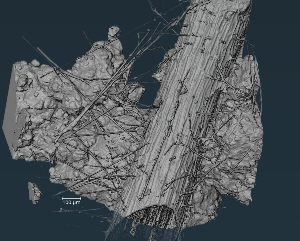
- Stacking the scans produced 3D reconstructions in which a root, thin hyphal filaments and surrounding sand grains appear together in place.
Compiled by The ScientistSomething wrong?How this is made
Why it matters
- capability Researchers can now look at the contact between living hyphae and mineral grains in undisturbed soil, the interface Stewart links to how fungal carbon reaches soil minerals.
- constraint With only a few dozen synchrotrons in existence, beamtime limits how many soils and plants the method can cover, so it suits targeted studies more than broad surveys.
- constraint The 13 billion ton carbon figure still rests on earlier research.
During the hours a high-resolution lab CT can spend on one sample, fungi move and grow, and the image blurs [8]. A synchrotron, a particle accelerator that makes very intense beams of light, cuts the exposure down [20]. "A synchrotron produces a million times the energy that your lab CT produces," said Braunmiller, a soil ecologist and graduate student at the Technical University of Munich [7][9]. "So you don't want to stand in the way of the beam. But then you can do the same images in a couple of minutes." [9]
Researchers can watch hyphae grow in petri dishes or in soil chips, which are silicon wafers built to mimic soil structure, but those settings leave out much of what the rhizosphere contains [18]. Imaging hyphae from real soil usually means extracting them first. Extraction leaves scientists without a 3D view of how roots, hyphae and spores connect [5]. Reporting in New Phytologist, Braunmiller and colleagues describe their approach as the first way to study the network in its natural environment [6]. Natural, in this case, means intact soil around living roots in a pot [11].
Each cylinder holds only about 6.8 cubic centimeters of soil [1], and a single gram of soil can contain up to 90 meters of mycelium [3]. The threads are about one tenth to one hundredth the width of a human hair [2]. When the team paged through the scans, the first sign of them was tiny white points moving across successive images [14]. "It was so exciting, because nobody was expecting that we could see them, because they're so tiny," Braunmiller said. "They're right at resolution edge." [13] Any hyphae finer than that edge would not appear in the reconstructions [13].
More than 80% of plants on Earth partner with mycorrhizal fungi to pull nutrients from soil [1]. Eos attributes to "some research" the suggestion that these fungi absorb the equivalent of 13 billion tons of carbon dioxide a year [4]. "Basically everyone on Earth is dependent on this really, really tiny zone around roots, in a way," Braunmiller said [19].
Justin Stewart, an ecologist with the Society for the Protection of Underground Networks and Vrije Universiteit Amsterdam, told Eos by email that studying hyphae within intact soils at higher resolution is a "major advance" [16]. "This makes it possible to examine the interface between living fungal networks and the mineral soil matrix, and opens new avenues for studying how fungal-derived carbon is transferred to soil minerals," he wrote [17].
I think Stewart's framing puts the claims in the right order. The scans show where hyphae meet mineral grains [15][17]. Measuring how much carbon crosses at those contacts is a separate experiment. The Eos account does not report the scan resolution, the share of hyphae the scans detect, or any carbon measurement.
What to watch
- Whether the New Phytologist paper reports a scan resolution and how much of each sample's hyphal network the scans actually detect.
- Follow-up work that pairs these scans with direct measurements of fungal carbon moving into soil minerals, the use Stewart points to.
- Whether the method is applied to field-collected soil cores or to plants other than tomato.