Science2 publishers3 min readPublished
Sleep debt gets a wiring diagram: mouse neurons that add and erase recovery sleep
Basel researchers mapped 162 mouse brains to find wake-activated cells in the median raphe and preoptic area. Switching them off cut sleep by nearly 70 per cent, with no rebound.
The Scientist · Science desk
Drafted by a language model from the sources cited here and checked against its claim ledger before publication. How we use AISend a correction
What happened
- By comparing whole-brain responses to sleep deprivation, recovery sleep and circadian behaviour in mice, researchers identified the anterior medial preoptic area and the median raphe as candidate regions that encode sleep deficit.
- The study used whole-brain activity mapping, targeted neuronal manipulations and electrophysiology to identify wake-activated neurons that regulate sleep drive in mice.
- Activating sleep-deprivation-responsive cells in the anterior medial preoptic area and median raphe induces increases in sleep duration and intensity that resemble recovery sleep.
- Inhibiting deprivation-responsive cells reduces sleep and abolishes the increased sleep propensity usually observed during deprivation.
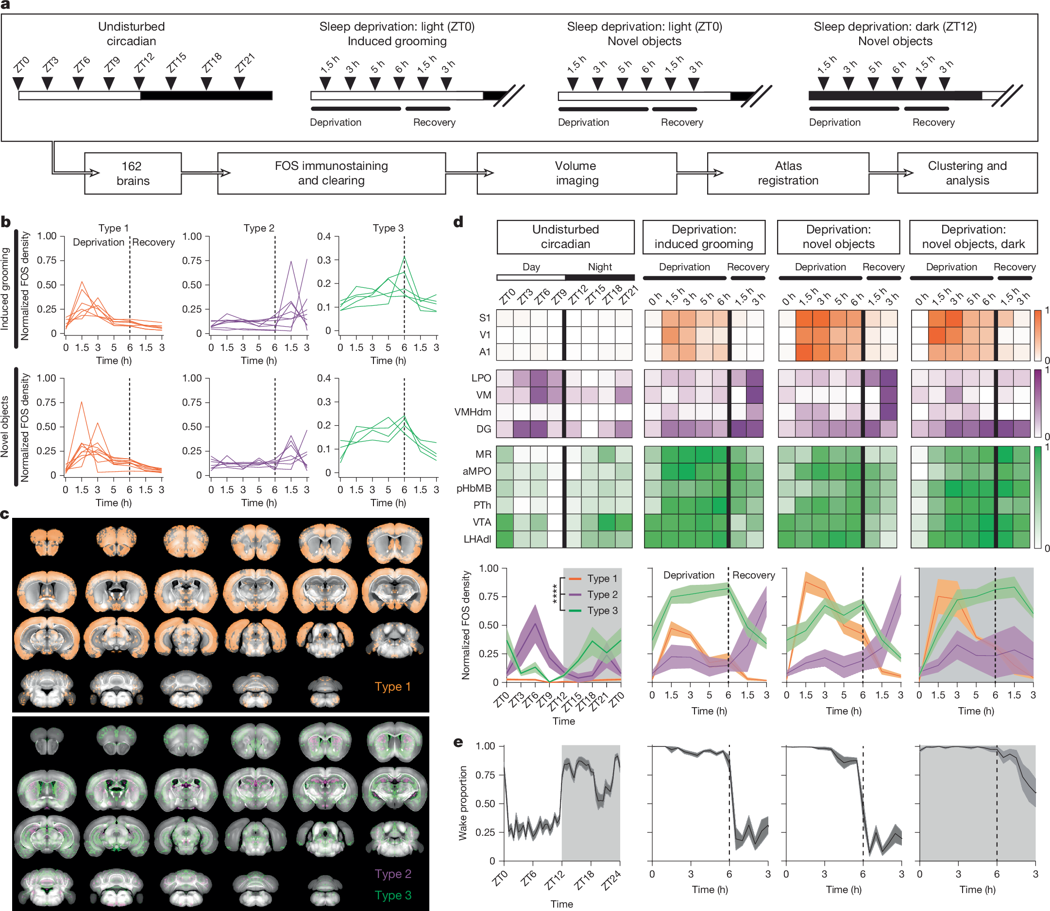
- Mice were deprived of sleep for 6 hours at the beginning of the light phase, their rest phase, using either induced grooming behaviour or novel object exposure.
Compiled by The ScientistSomething wrong?How this is made
Why it matters
A group at the University of Basel has identified wake-activated neurons in mice that encode accumulated sleep deficit, and has shown that turning them on produces something resembling recovery sleep while turning them off erases the urge to catch up [1][2][3][4]. Homeostatic sleep drive has been a quantity inferred from behaviour and EEG for decades; this makes it a set of addressable cells.
The mapping came first. Mice were kept awake for six hours at the start of the light phase, their rest phase, using induced grooming or exposure to novel objects [5]. Brains were then stained for FOS, a marker of recently activated neurons, cleared, and imaged by light sheet at four points during deprivation and two during a three-hour recovery window [6]. Dark-phase deprivation and ordinary three-hour circadian intervals served as controls for stimulus-specific and time-of-day effects [7]. Across 162 brains and 26 conditions, voxel-wise clustering separated three response profiles: an early-peaking type that tracks the onset of the deprivation stimulus and peaks 1.5 to 3 hours in, a sleep-correlated type, and a wake-correlated type that scales with time awake [8][9]. The anterior medial preoptic area and the median raphe fell into the third category [1].
Then the interventions. Activating the deprivation-responsive cells in those regions increased both sleep duration and sleep intensity, the latter measured as delta-band EEG power around 0.5 to 4 Hz [3][10]. According to New Scientist, mice with both median raphe populations activated slept two to three times as long as controls and spent more time in non-REM slow-wave sleep [11]. Inhibition ran the other way: less sleep, and, critically, no rise in sleep propensity during deprivation at all [4]. The deficit signal was not merely muted; it was not produced.
The responsive cells in the median raphe include serotonergic neurons and a separate GABAergic population whose intrinsic excitability rises during deprivation [12]. They project to subcortical sleep-associated regions and act through the preoptic hypothalamus [13]. Co-activating both types promotes sleep synergistically; co-inhibiting both chronically cuts sleep by nearly 70 per cent [14].
That last figure carries the strangest result. Chronically inhibited mice lost about 6.5 hours of sleep per day, implying a baseline near 9.3 hours and a residual under three [15][16]. The Nature authors report that most animals survived without the compensatory sleep drive or behavioural deficits that severe deprivation normally causes [17], and New Scientist reports no increase in the anxiety-like behaviours mice usually show [15]. But roughly 17 per cent of the mice died, meaning about 83 per cent survived [18][19]. "Most survive" and "one in six does not" are both true statements about the same experiment, and the second one constrains any therapeutic reading of the first.
Two caveats are load-bearing. Whether the equivalent populations exist in humans is unknown, and the researchers say therapy would depend on first working out how the cells talk to other regions [20]. And this is one path among several: Mark Wu at Johns Hopkins, whose group has implicated the thalamic nucleus reuniens in tracking the gap between sleep needed and sleep obtained, places the work in a growing set of wake-activated, sleep-promoting circuits [21], while Luis de Lecea at Stanford notes that the brain has many ways to fall asleep and wake up [22]. Chiara Cirelli at Wisconsin-Madison called the whole-brain FOS mapping a tour de force [23].
What to watch: whether the 70 per cent reduction holds without rebound over longer horizons, what killed the 17 per cent, and whether the GABAergic excitability shift has any measurable human correlate.