Science1 publisher2 min readPublished
Two RAB1 proteins bridge the adaptor OPTN to the vesicles that seed mitophagy membranes
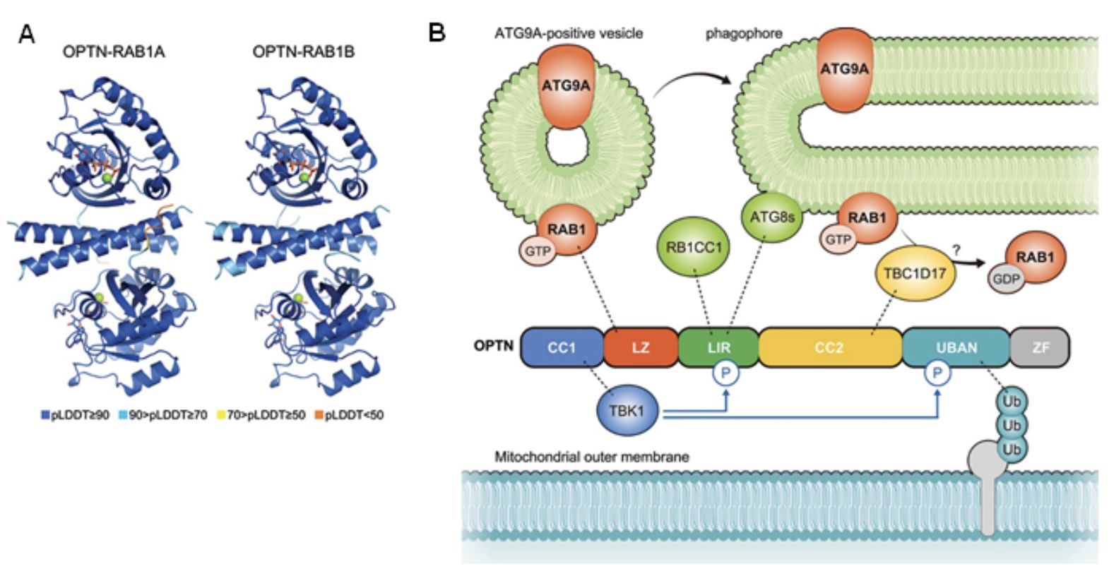
Tokyo Metropolitan Institute of Medical Science researchers report that RAB1A and RAB1B, two of about 60 human RAB proteins, tie OPTN to ATG9A vesicles. The link helps set where the membrane around a damaged mitochondrion begins to form, and labs can now test that step directly.
The Scientist · Science desk
Drafted by a language model from the sources cited here and checked against its claim ledger before publication. How we use AISend a correction
What happened
- The same group had already shown that OPTN pulls ATG9A vesicles, thought to supply an autophagosome's membrane seed, to damaged mitochondria, but how it did so was unknown.
- Silencing RAB1A and RAB1B together with RNA interference left ATG9A vesicles unable to gather efficiently at OPTN sites.
- Suppressing RAB1 also reduced mitophagy driven by the PINK1-Parkin pathway.
- RAB1 attaches to ATG9A vesicles through prenylation at its C-terminal end.
Compiled by The ScientistSomething wrong?How this is made
Why it matters
- capability Both ends of the bridge are now mapped, so the OPTN-RAB1 contact can be broken selectively in disease-relevant cells to ask whether clearing damaged mitochondria depends on it.
- constraint Because RAB1A and RAB1B were silenced together, the data cannot yet assign the job to either one, so a follow-up that targets a single RAB1 gene starts without evidence it would be enough.
- precedent The result strengthens the group's case that adaptors such as OPTN recruit the membrane that will engulf their cargo, an idea other ubiquitin-reading adaptors can now be checked against.
RAB proteins were the obvious place to look for the missing connector. Humans carry about 60 of these small GTPases, and they work as switches that regulate membrane traffic inside the cell [6]. The Intracellular Quality Control Project at the Tokyo Metropolitan Institute of Medical Science screened RAB proteins for binding to OPTN, and RAB1A and RAB1B came back [1][6]. The study is published in Autophagy [3].
A hit in a binding screen shows that two proteins end up together. It does not show that they touch. The group predicted the interface with AlphaFold, ran an interaction assay inside cells, and then broke the contact with mutations [9]. I like the sequence: the prediction proposes a contact, and the mutations test whether it is real. The leucine zipper is a separate domain from UBAN, the part of OPTN that recognizes ubiquitin chains [11]. In the group's model, OPTN reads the ubiquitin on a damaged mitochondrion and binds RAB1 at the same time, and RAB1 brings an ATG9A vesicle with it [12].
The authors conclude that RAB1 plays an essential role in connecting OPTN to the trafficking machinery that builds the autophagic membrane [13]. The thing this doesn't tell you is how much of the drop in mitophagy [8] runs through OPTN in particular. RAB1 is a general trafficking switch [6]. Depleting it could slow mitophagy through other steps as well. The cleaner test is to put back an OPTN whose leucine zipper cannot bind RAB1 and see whether mitophagy fails. The published summary does not say whether that experiment was run, or how large the reduction in mitophagy was.
PINK1 and Parkin build the ubiquitin signal on damaged mitochondria that OPTN reads [4]. This study adds the step after that signal: placing the membrane seed at the organelle that has to be removed [2]. The work as reported examines the axis during PINK1-Parkin mitophagy [14]. Tying it to a specific disease where mitochondrial cleanup fails will take patient cells or animal models.
What to watch
- Whether an OPTN leucine-zipper mutant that cannot bind RAB1 fails to support mitophagy when put back into cells.
- Whether RAB1A or RAB1B alone is sufficient to recruit ATG9A vesicles to OPTN sites.
- Tests of the OPTN-RAB1-ATG9A axis in patient-derived cells or animal models where damaged mitochondria accumulate.