Science1 publisher3 min readPublished
Carotid arteries on a chip turn stroke prevention into a test you run, not a guess
Sydney researchers 3D-printed six patients' diseased carotids, lined them with cells and flowed human blood through to watch clots form and shed. It is proof-of-concept, aimed at recurrent-event cases.
The Scientist · Science desk
Drafted by a language model from the sources cited here and checked against its claim ledger before publication. How we use AISend a correction
What happened
- Researchers pioneered a way to create miniaturized copies of a person's carotid artery that could help doctors predict and manage the patient's stroke risk; the study was published in July in the journal Cell Biomaterials.
- Scientists used the 'arteries-on-a-chip' to monitor how real blood flows through a patient's carotid arteries, which carry blood to the brain, face and neck.
- The approach could enable doctors to identify not just how and what type of clots form in that specific patient, but also determine which medications would be most effective in dealing with the blockage.
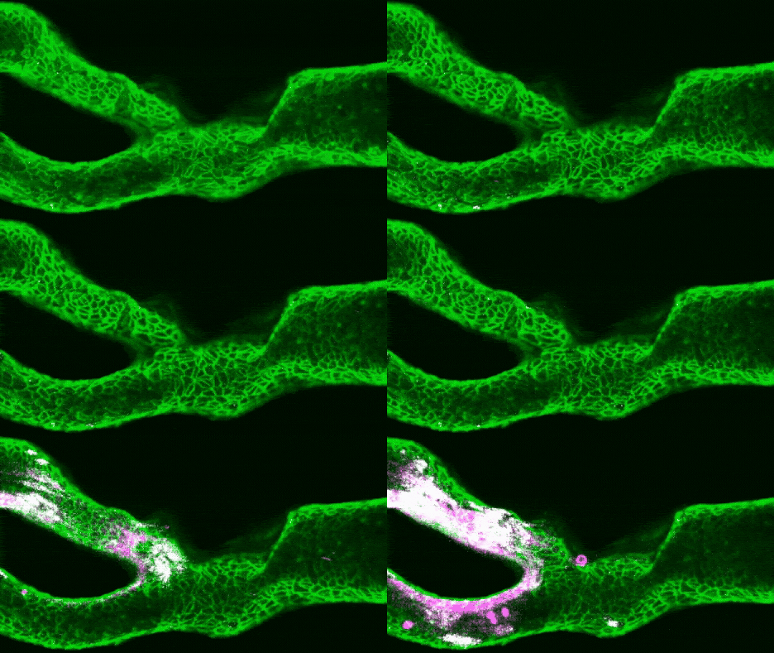
- To build the models, the team used a patient's existing CT scans to 3D print a plastic replica of their carotid artery including any narrowing caused by atherosclerosis; the inside was coated with collagen, then cells that line the carotid artery were layered on top, and blood was passed through mimicking the speed and pressure of blood flow in the body.
- Zhao: 'Then [we] use a very small laser injury to expose the collagen underneath. We can then flow human blood through the artery and watch thrombosis happen under a microscope.'
Compiled by The ScientistSomething wrong?How this is made
Why it matters
Researchers have built miniature, cell-lined replicas of individual patients' carotid arteries and flowed real human blood through them to watch clots form and break apart, in work published in July in the journal Cell Biomaterials [1][2]. The interesting part is not the printing; it is the reframing of stroke prevention as an assay you run on a specific patient's vessel and blood, rather than a therapy chosen from population averages [3].
The build is deliberately unglamorous. The team starts from a patient's existing CT scans, 3D prints a plastic replica of the carotid including whatever narrowing atherosclerosis has produced, coats the inside with collagen, layers on the cells that line the carotid, then pushes blood through at the speed and pressure of the real circulation [4]. A small laser injury exposes the collagen underneath, and thrombosis is then watched under a microscope as human blood flows past [5]. That injury step matters mechanistically: according to first author Charles Zhao, a doctoral student at the University of Sydney, damage to the artery lining exposes collagen, von Willebrand factor grabs platelets out of the flow, and those platelets stick together and recruit more [6][7].
The clinical gap the model targets is the difference between anatomy and behaviour. "Clinically, we are very good at imaging how narrow an artery is," Zhao said, "but narrowing alone does not tell us exactly how a clot will behave" [8]. A clot can sit firmly on the wall, mildly impeding flow and posing no immediate danger, or shed fragments that travel toward the brain and block smaller vessels [9]. Across six patients with different types of arterial damage, combined with flow calculations, the team reports a more accurate picture of clot formation and shedding than imaging alone gives [10][11]. Two arteries with apparently similar disease behaved very differently, Zhao said, because their three-dimensional shapes created different flow patterns, and those local conditions determined whether a clot grew stably or shed fragments [12].
That is the drug-selection argument. Some treatments work by preventing platelets from clumping into clots [13], so a bench readout of whether a given patient's clot is a stable mound or a fragmenting one is functional information that a stenosis percentage cannot supply. Zhao puts the use case narrowly: "patients who have experienced recurrent events despite treatment, or cases where clinicians have several possible treatment strategies but limited functional information to distinguish between them" [14]. That is the right claim to make, and it is a small population by design.
Scale check. Ischemic stroke is described as a leading cause of death worldwide, second only to heart disease, and roughly one in five of these events results from atherosclerosis [15][16] - meaning about four in five do not, and sit outside what a plaque-shaped carotid replica can model [17].
What to watch: whether the chip's prediction of clot stability tracks what actually happened to those patients, and whether a drug chosen on the chip outperforms one chosen conventionally. The published account is explicitly proof-of-concept and reports no turnaround time, no cost, and no comparison against clinical outcomes [18]. Six vessels, one laser injury model and no outcome data is a long way from a susceptibility test a stroke unit can order. The path from here runs through blinded prediction against known follow-up, then a trial where the assay changes the prescription.