Science1 publisher3 min readPublished
A chip with human bone, lung and circulating tumour cells tests the metastasis model gap
Columbia researchers report a multi-organ chip in which breast cancer cells in vascular flow colonise engineered bone and lung differently, matching known organ tropism.
The Scientist · Science desk
Drafted by a language model from the sources cited here and checked against its claim ledger before publication. How we use AISend a correction
What happened
- Cancer metastasis is responsible for at least two-thirds of cancer deaths.
- Drugs targeting metastatic progression have largely failed, in part due to the lack of predictive models that would help identify the underlying mechanisms of metastasis.
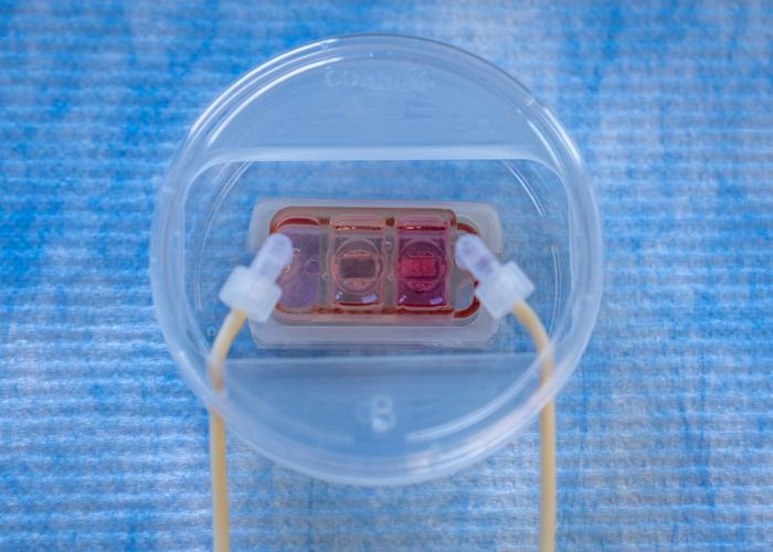
- New work reports development of a multi-organ chip that mimics how cancer cells spread from vascular flow to distant organs, described as the first model of cancer metastasis of its kind.
- The chip includes compartments with millimeter-sized engineered human bone and lung tissues, plus vascular flow containing circulating breast cancer cells, allowing dynamic cross-talk of the tissues being colonized.
- The work is published in Science Translational Medicine in the paper 'Organ-specific colonization and niche remodeling in a human tissue model of metastasis.'
Compiled by The ScientistSomething wrong?How this is made
Why it matters
Researchers at Columbia University have reported a multi-organ chip in which engineered human bone and lung tissues sit in separate compartments linked by a vascular circulation carrying breast cancer cells, described as the first model of cancer metastasis of its kind [3][4]. The reason to care is arithmetic rather than novelty: metastasis is responsible for at least two-thirds of cancer deaths, and drugs aimed at metastatic progression have largely failed, in part because there are no predictive models to identify the mechanisms those drugs would have to interrupt [1][2].
The work appears in Science Translational Medicine as "Organ-specific colonization and niche remodeling in a human tissue model of metastasis" [5]. The bone, the lung and the vascular endothelium were all engineered from induced pluripotent stem cells using tissue-specific scaffold-bioreactor culture systems, with bone and lung chosen because they are common sites of breast cancer metastasis [10]. Each millimeter-sized tissue sits in its own compartment, optimised for maturation and long-term maintenance of function, and the compartments are joined by vascular circulation [4][11]. A selectively permeable endothelial barrier separates each tissue from the vascular channel, as in the body [12].
The load-bearing result is that the two tissues did not behave identically. Cancer cells that typically gravitate toward bone showed stronger bone colonisation and induced more pronounced bone degeneration, while cells that typically gravitate toward lung caused greater disruption in lung tissue and only modest bone colonisation [13][14]. According to the authors, those distinct patterns of colonisation and of secreted factors show the device reproduces key features of organ-specific metastasis seen in patients [15]. Post-analysis of the engineered tissue also showed cancer cells conditioning the distant organs to be more receptive before colonising them, with signs of pre-metastatic niche formation in both compartments [16].
Gordana Vunjak-Novakovic, a university professor and professor of biomedical engineering and of medical sciences at Columbia, said the key advantages are that the model is human and can be patient-specific, and that it mimics aspects of human metastasis that are otherwise largely inaccessible to direct study [6][7]. She framed the objective as probing whether cancer cells can adhere to and cross the endothelium, and whether they survive in the tissues they colonise through cell reprogramming and niche remodeling [8]. "Cancer is very smart, unfortunately," she said, adding that the team learned how cells cross barriers out of circulation and reproduced the conditioning of target tissues seen in patients [17]. Colonisation is precisely the phase that animal models handle badly, since it requires cancer cells to evade tissue defences and adapt to a specific organ [9].
Two limits are worth holding onto. Patient specificity is stated as a property the platform can have, not as a reported experiment with patient-derived cells [20]. And the account of the study contains no drug-response or drug-screening result, which is the test that would show whether the chip predicts anything a mouse does not [19].
What to watch: whether the secreted-factor signatures hold up when the cells come from individual patients rather than tropism-selected lines, and whether the platform is used to rank candidate compounds. The report situates the study within the FDA's and NIH's growing emphasis on new approach methodologies, which is where any regulatory weight would come from [18].