Science1 publisher3 min readPublished
One hand-annotated membrane trains MemBrain v2 to pick protein complexes on the rest
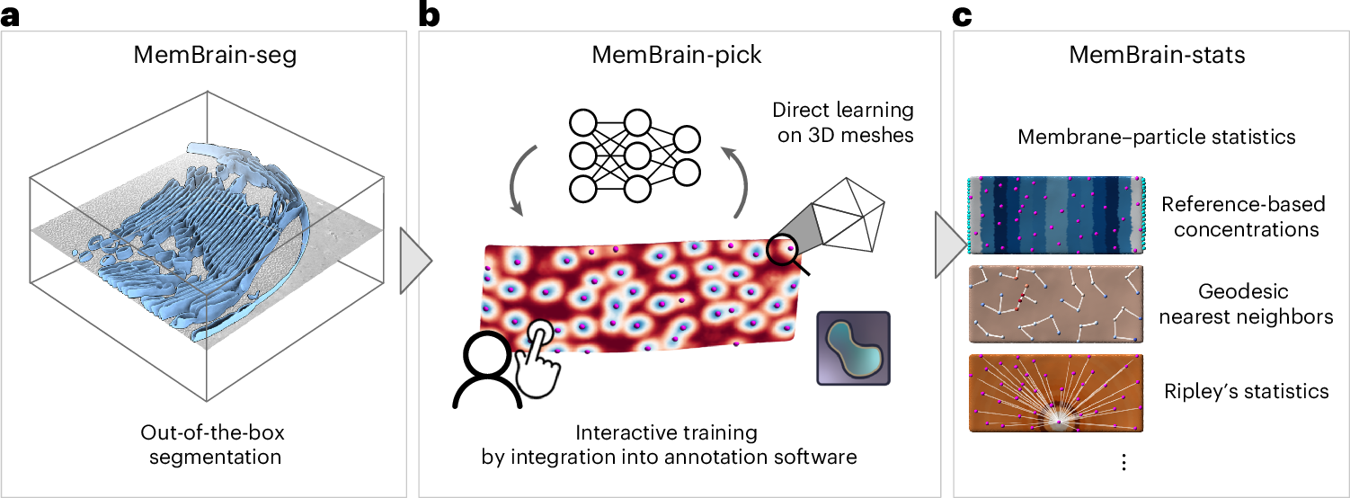
MemBrain v2, published in Nature Methods by groups in Munich and Basel, reports a 91% F1 score locating membrane protein complexes on new membranes after a single one was labelled by hand. Its authors put the time saving at weeks down to a few hours.
The Scientist · Science desk
What happened
- Helmholtz Munich, the Technical University of Munich and the Biozentrum of the University of Basel published MemBrain v2 in Nature Methods, an AI tool for analysing membranes in 3D images of cells.
- In one test the team hand-annotated the positions of protein complexes on a single membrane, and the pick module then located the same complexes on additional membranes with an F1 score of 91%.
- The group reports that analysis work which once took weeks of manual labelling now takes a few hours with the software, which is freely available.
- All components are open source, and the membrane-detection module is already in use internationally, including across data sets from the Chan Zuckerberg Imaging Institute.
Compiled by The ScientistSomething wrong?How this is made
Why it matters
- capability Seeding the pick module from one hand-annotated membrane means the annotation cost of a new project scales with the number of distinct sample types, not with the number of tomograms collected.
- constraint Every hour saved lands after data collection, so a lab whose ceiling is microscope access or sample preparation gains analysis capacity it has nothing to feed.
- decision At 91% F1 someone still has to check the picks, so a lab re-planning headcount is moving annotation hours into validation hours.
- precedent With the code open, a skeptical reviewer can rerun segmentation on the same tomograms. That raises the bar for any membrane analysis published on hand-drawn labels alone.
An F1 of 91% is one number covering two different mistakes [8]. Because F1 is the harmonic mean of precision and recall, a precision of 1.0 at that score implies a recall of 0.835, so neither figure can be worse than about 84% [17]. Inside that band the behaviour varies a lot. The pick module could be missing roughly one complex in six while flagging almost nothing spurious, or doing the reverse, and a combined score does not separate the two [18].
Which one dominates matters for the third module, because MemBrain-stats measures how the picked proteins are arranged [10]. The gaps in cryo-ET data are not random with respect to geometry. "One challenge is that cryo-ET images can contain gaps in information due to technical limitations of the imaging process. As a result, certain membrane orientations are difficult or partly impossible to see. This is exactly where MemBrain v2 comes in, automating the process," said first author Lorenz Lamm [6]. In my view a miss concentrated on those orientations would bias a spacing or clustering readout.
The weeks-to-hours figure is the team's own summary of the gain [2], and the paper does not say how many tomograms were involved on either side of the comparison [19]. In several applications, the authors report, the tool matched the results of painstaking manual analyses and was considerably faster [11]. Before it, hand labelling rarely transferred to a new data set, and available programs generally covered one part of the job, either outlining the membranes or locating the proteins inside them [9]. MemBrain v2 puts membrane finding, protein picking and arrangement statistics in a single tool [10].
The segmentation module needs no annotations or training data from the user at all, and the pick module needed one membrane [7]. Depending on the application, the tool as a whole needs little or no extra training data [4]. "By making these analyses faster and accessible to research groups worldwide, we can study cellular processes across much larger data sets," said senior author Tingying Peng [13].
For a lab planning its year, only part of the pipeline gets cheaper. Cryo-ET still requires flash-frozen cells and a collected 3D data set before any of this runs [5], and MemBrain v2 starts at membrane detection [10]. If microscope access or sample preparation sets the ceiling, a faster analysis step does not raise tomograms per month.
The tool has already been used in a separate study that found photosynthesis proteins spatially separated within the membrane, against previous models of their organization [14]. The authors say it is set to distinguish different protein types more precisely in future [15]. The paper is Lamm et al, Nature Methods (2026), DOI 10.1038/s41592-026-03178-8 [16].
What to watch
- An independent group reporting precision and recall separately, on samples the Munich and Basel authors did not use, would settle how the residual 9% divides.
- Whether MemBrain-pick transfers between different sample types, not only between membranes within one data set.
- Whether the promised finer discrimination between protein types arrives, since that is what the authors say comes next.