Science1 distinct publisher3 min readPublished
Spatial-ATAC-Hi-C pulls contact maps and accessibility signal from one barcoded spot of mouse and human brain tissue, which is how its authors come to report copy number and structural variants region by region across glioblastoma and astrocytoma.
The Scientist · Science desk

Compiled by The ScientistSomething wrong?How this is made
3C-based methods including Hi-C exist to expose the hierarchy of nuclear architecture: compartments, topologically associating domains, chromatin loops [7]. Accessibility assays such as spatial-ATAC-seq report which regions are open, and spatial-CUT&Tag adds histone marks like H3K27ac and H3K27me3 [5]. Run those on adjacent sections and you align them by coordinate and trust that two slices were the same tissue. Take them off the same barcoded pixel, which is what this platform is described as doing [1], and the pairing stops being an assumption.
The multi-omics stack the authors survey combines epigenome, transcriptome and proteome [8]. The nearest prior attempt at spatial genome folding is Spatial-SPRITE, which they describe as microfluidics-based and as something that "potentially can generate spatially mapped SPRITE data" [9]. The word "potentially" is theirs, and it marks how young this corner of the field is.
The pixel is the detail the abstract skips over. The same introduction puts spatial transcriptomics resolutions between 55 micrometres and 2 micrometres, some reaching single-cell or subcellular scale [10]. Those endpoints are 27.5-fold apart in linear dimension and about 756-fold apart in area [11], and area is what decides how much DNA sits under one barcode. So the first questions are how many usable contacts land in a pixel, at what pixel size, and how deep the sequencing had to run to get them. Those figures, and the sample counts, are simply absent from the material we have [12].
In the tumour sections the ambition is diagnostic rather than demonstrative: 3D genome alterations, copy number variations and structural variations mapped across regions [3], read by the authors as clinically relevant oncogenic events and clonal heterogeneity [4]. Slide-DNA-seq already placed copy number alterations of tumour clones in spatial context [6], and single-cell 3C methods already showed cell-to-cell heterogeneity in higher-order structure [14]. What is new is the joint readout: a folding change and an accessibility state from one barcode, so a lab can ask whether a structural variant sits beside newly opened regulation. Which change came first is a separate question from co-location, since both most likely sit downstream of whatever the clone has been doing to its own genome, and the authors' own framing puts 3D organisation upstream of gene regulation in development, immune response and disease progression [15], which is a reason to look, not a licence to infer direction.
One caveat this paper is spared: the brain work covers human as well as mouse tissue [2]. Whether it survives contact with a pathology service depends on throughput and cost per slide. The full paper, titled "Spatial chromatin architecture and accessibility co-profiling of mammalian tissues" [13], presumably reports the yield figures; the abstract, which is what most people will read, carries none of them [12].
Ranked by verification strength, evidence, and original report placement.
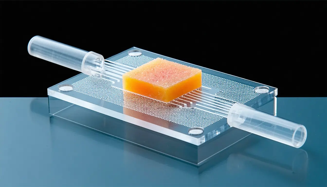
Spatial-ATAC-Hi-C is a microfluidic-based platform for genome-wide, spatially resolved joint profiling of 3D genome organisation and chromatin accessibility on tissue slides.
Applied to mouse and human brains, Spatial-ATAC-Hi-C revealed distinct chromatin architecture and gene regulatory programs in neuronal and non-neuronal populations in their native tissue context.
In glioblastoma and astrocytoma samples, the authors detected spatially resolved 3D genome alterations, copy number variations and structural variations across tumour regions.
Spatial epigenomic techniques such as spatial-ATAC-seq and spatial-CUT&Tag allow spatial profiling of chromatin accessibility and histone modifications (H3K27ac and H3K27me3) within local tissue regions.
Slide-DNA-seq provides an opportunity to interrogate the copy number alterations of tumour clones within their spatial tissue context.
Hi-C and other 3C-based techniques have frequently been used to reveal hierarchical features of nuclear architecture, including compartments, topologically associating domains and chromatin loops.
Distinct publishers with included, body-backed reporting in this cluster.
1 article · August 31, 2026
Follow any of these and your For You feed starts watching them — no settings page required.
science
REDCAT reads metabolic state and cell identity off one section, in ratios not molecules1 distinct publisher
product
Vivodyne says the AI drug bottleneck is human tissue data, not model capability1 distinct publisher
Evidence-backed comparisons of source perspectives and observed adoption signals. Read the methodology
Which Builder, Operator, and Investor concerns the observed source mix emphasized—not a truth score.
Evidence, demonstrated adoption, hype gap, incentives, and confidence are assessed independently, each on its own current evidence. How these are measured.
Peer-reviewed, single-voiced, truncated
A Nature Methods paper is a solid provenance floor, and the field context — Hi-C compartments and TADs, spatial-ATAC-seq, slide-DNA-seq — is standard and checkable. But the claims that matter live in an abstract, and the supporting text breaks off at the word 'Develop' where Results should begin. What we can verify is that the method is described; what we cannot see is the data behind neuronal versus non-neuronal architecture or the tumour variant calls.
Publication is the only event
Nothing has happened yet except the paper appearing. No second laboratory has run the protocol, no vendor has picked up the chip, no dataset release or clinical pipeline is mentioned anywhere in this reporting. Scoring uptake from the inventors' own demonstration tissues would be inventing a number.
Clinical adjectives, unhedged
Notice the asymmetry in the authors' own prose: a competitor's Spatial-SPRITE 'potentially can' generate spatially mapped data, while their own tumour work arrives already 'revealing clinically relevant oncogenic events' and the platform is already 'a powerful tool'. The overstatement is modest and mostly rhetorical — the method itself is described soberly — but a paper that quotes 55-to-2-micrometre resolutions for neighbouring techniques and never states its own is asking to be taken more on trust than the visible text earns.
The builders are the only narrators
Every sentence in our coverage was written by the people who invented the instrument, in the venue where methods papers earn citations and follow-on funding. The introduction is structured as a gap argument: imaging methods cover 'selected loci or regions', prior spatial assays cover one layer, single-cell 3D genome work loses tissue context — therefore this platform. That is normal scientific persuasion, not misconduct, but it is the only voice in the room and it has a stake in the conclusion.
Believe the method, not the magnitudes
Two things are firm: this platform exists and it was published in a journal that reviews methods seriously. Almost everything a reader would want to do with that — judge resolution, compare against droplet Hi-C or slide-DNA-seq, assess whether the glioma clones are convincingly separated — depends on numbers not present and on replication not yet attempted. Confidence sits mid-scale and is capped by the single-source structure rather than by any contradiction.