Science1 publisher2 min readPublished
Copenhagen traces some heart defects to a signalling hub inside the cell's antenna
A University of Copenhagen team paired rare-variant data from several thousand heart-defect patients with engineered zebrafish and cell experiments. The brain, kidney and skeletal effects in its summary are predictions.
The Scientist · Science desk
What happened
- A University of Copenhagen group reports that three proteins, TAK1, TAB2 and PKA-C-alpha, act together as a signalling hub inside the primary cilium, and that their activity is important for normal heart formation.
- The work started with genetic data from several thousand patients with congenital heart defects, looking for rare variants that appeared more often in patients than in healthy individuals.
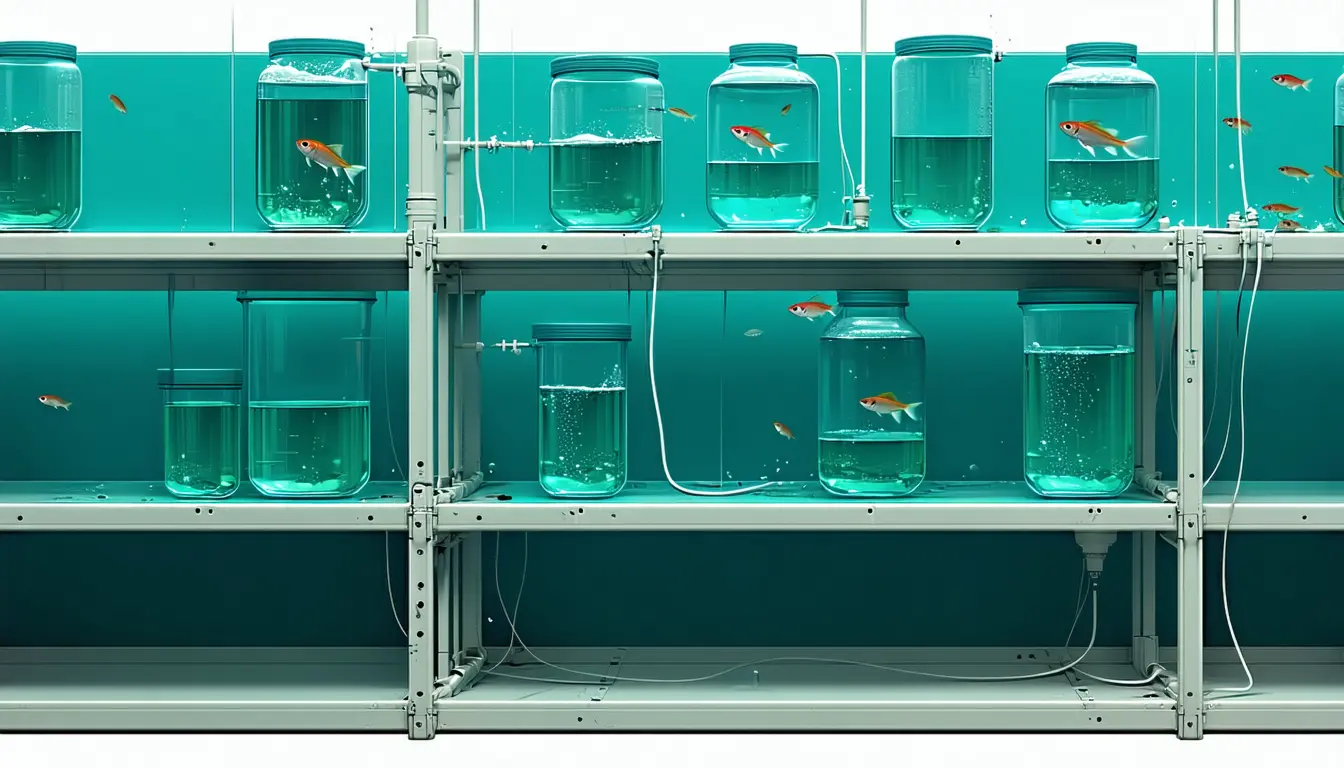
- The team then engineered those same mutations into zebrafish, and reported that the gene changes interfered with normal heart development and reduced heart function in the fish.
- The study addresses syndromic congenital heart disease, the form that arrives as part of a wider genetic syndrome with abnormalities elsewhere in the body.
Compiled by The ScientistSomething wrong?How this is made
Why it matters
- constraint The causal test lives in zebrafish and cultured cells, so reading this as the explanation for a particular child's heart defect crosses a species boundary the experiments did not cross.
- capability Because cilia sit on most cell types, clinicians get a checkable expectation: some syndromic heart patients should carry kidney, brain or skeletal findings traceable to the same variant.
- decision Genetics labs now have a specific question to put to their own cohorts, namely whether cilium-pathway variants are enriched enough in syndromic heart-defect patients to earn a place on clinical sequencing panels.
A variant-burden comparison ranks genes. It cannot show what a variant does to a forming heart. That is what the fish are for: engineering the same mutations into zebrafish is the step that tests causation, and it is also the step furthest from a patient [8][9]. The laboratory work sat in between, in human cells and mouse stem cells, and was used to trace the signalling itself [10].
The three proteins are TAK1, TAB2 and PKA-C-alpha. The claim is that they work as a hub inside the primary cilium, the antenna-shaped structure that extends from the surface of most cells in the body [4][2]. Cilia are how a cell reads chemical signals from its surroundings, including signals that bear on whether it divides, moves or dies [3]. "This finding changes our understanding of why some congenital heart defects arise," said Lars Allan Larsen, professor at the Department of Cellular and Molecular Medicine. He added: "You could say that we have identified an important cog in a highly complex machine." [6][5]
The University of Copenhagen release gives no figures for the zebrafish result, and none for the variant frequencies behind the human comparison [17]. So the human side of the case remains an association between how often a rare change appears in patients and how often it appears in healthy people [8].
The summary goes further than the heart, saying that when mutations disrupt this system the effects may extend beyond it to organs including the brain, kidneys and skeleton [12]. That expectation follows from where cilia are, since they sit on most cell types [2], while the experiments described are cardiac [9].
The epidemiology quoted in the release comes from the World Heart Federation and the Danish Heart Foundation. Roughly two in every 100 newborns worldwide, 2.3 to 2.5 million affected births a year, about 16 million people living with congenital heart disease as of 2023 [13][14]. Divide 16 million by 2.4 million and the prevalent population equals 6.7 years of births [15]. People born with heart defects accumulate over decades, so those two figures are counting different populations, and neither is broken out for the syndromic cases this study addresses [14][11].
"These proteins act as molecular instructions that tell stem cells when and how to develop into heart muscle cells," said Soren Tvorup Christensen, professor of cell biology at the Department of Biology [7]. The stem cell experiments used mouse cells [10].
What to watch
- Publication of the underlying paper, with the size of the zebrafish cardiac deficit and the case and control counts behind the human variant comparison.
- Whether patients carrying TAK1, TAB2 or PKA-C-alpha variants are re-examined for kidney, brain and skeletal abnormalities.
- Whether an independent patient cohort reproduces the enrichment of these variants.