Science1 publisher3 min readPublished
Brain organoids build the right cells in the wrong order, and that dents size-disorder models
A Nature study reports that mouse organoids make cortical cell types but not the clock that sequences them, and that a third of progenitors get locked into a single fate.
The Scientist · Science desk
Drafted by a language model from the sources cited here and checked against its claim ledger before publication. How we use AISend a correction
What happened
- The brain develops in stages, and it is important that certain cells form before others so that precise neural connections can emerge later.
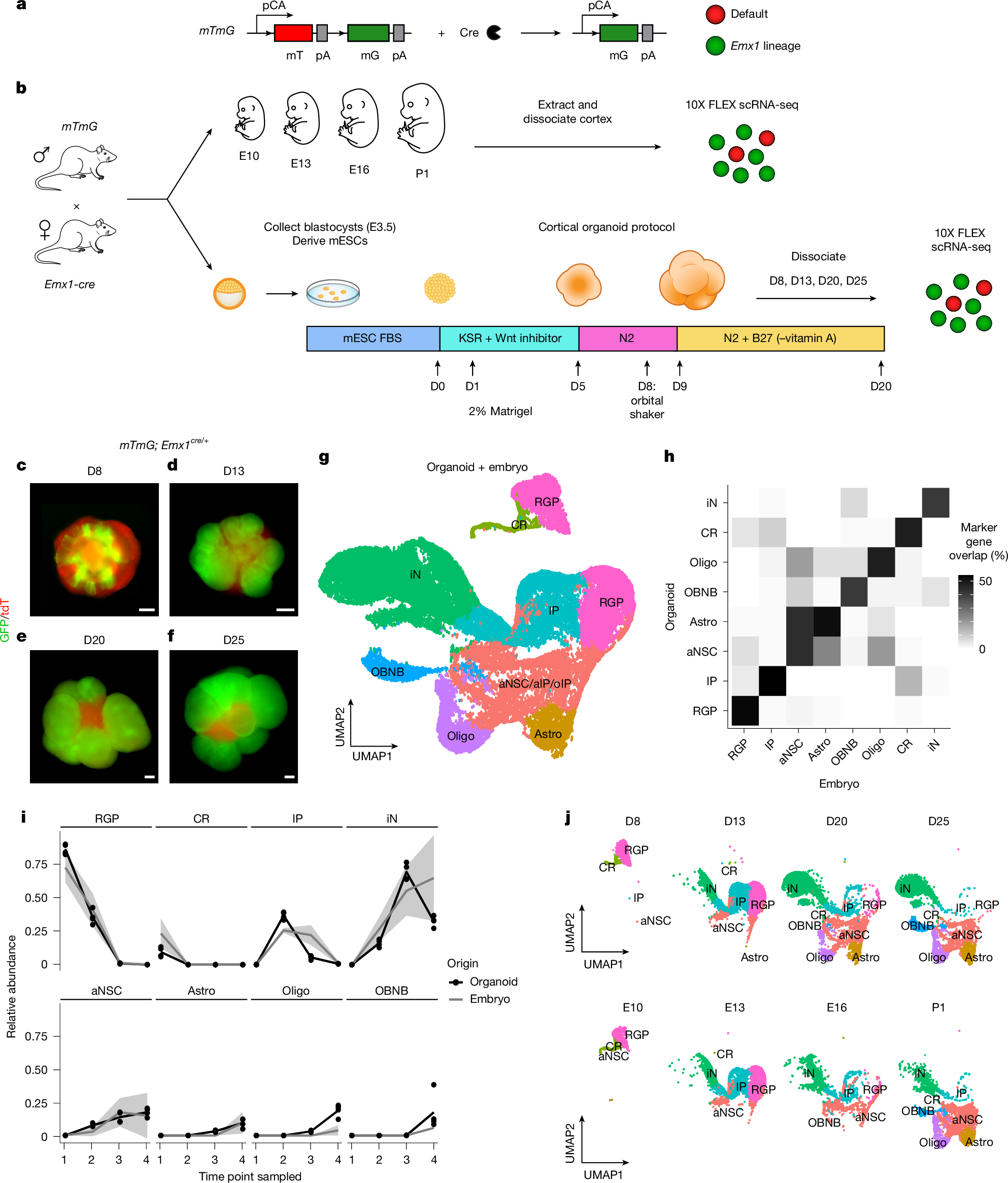
- A new study published Aug. 12 in the journal Nature suggests that brain organoids may be missing some crucial ingredients that would help them resemble real brains.
- Study co-author Simon Hippenmeyer, a neuroscientist at the Institute of Science and Technology Austria, said that if the precisely timed processes of brain development go wrong it can lead to conditions like macrocephaly and microcephaly, in which the brain grows abnormally large or small.
- Because it is not possible to directly study microcephaly and macrocephaly in human fetuses, scientists turn to brain organoids.
- In earlier mouse experiments, Hippenmeyer's team tracked radial glial progenitors (RGPs) during early embryonic development by giving pregnant mice a drug that activated fluorescent labels on individual stem cells, then traced the resulting lineages after birth to see how many cells they produced and how that number changed as development progressed.
Compiled by The ScientistSomething wrong?How this is made
Why it matters
A study published Aug. 12 in Nature reports that brain organoids grown from mouse embryonic stem cells produce the same major cell types as a real brain, but not in the same sequence [2][6][7]. The two conditions that organoid work in this area most often targets, microcephaly and macrocephaly, arise when precisely that timing goes wrong, according to study co-author Simon Hippenmeyer, a neuroscientist at the Institute of Science and Technology Austria [3].
Cortical development runs on an order of operations: certain cells have to form before others so that precise connections can emerge later [1]. Radial glial progenitors (RGPs) in the cerebral cortex first divide to expand their own numbers, then differentiate into neurons and the glial cells that support, nourish and insulate them [8]. Hippenmeyer's group had already measured how that plays out in live mice, giving pregnant animals a drug that switched on fluorescent labels in individual progenitors and then tracing the lineages those cells produced after birth [5]. That dataset became the yardstick for the organoids [6].
Three deviations came out of the comparison. The phases ran concurrently rather than in sequence: some organoid RGPs formed neurons early while others were still proliferating [9]. The output per progenitor lost its downward slope, which is the more consequential finding. In a real brain, RGPs at later stages yield fewer descendants than early ones; in the organoids they yielded more, regardless of developmental time frame [10]. And fate flexibility narrowed. A single RGP normally can make both deeper-layer neurons, earlier, and upper-layer neurons, later, across the cortex's six layers, but about a third of organoid RGPs were restricted to one fate and could not make the other type [11][12]. Roughly two in three kept both options [13].
Descendants per progenitor is close to the quantity that separates an abnormally small cortex from an abnormally large one, and in the organoids its developmental trend is inverted [10]. A system that mismeasures the variable under study is not a neutral instrument: a growth phenotype in such a dish could reflect the culture as easily as the mutation. That reading is mine, not the study's, but it is hard to avoid given that organoids are used here because human fetuses cannot be studied directly [4], which is also why the discrepancy is hard to check.
On why organoids drift, the researchers point to what the dish lacks. A living brain contains blood vessels, extracellular structures and metabolic signals, and the team thinks those external cues help coordinate the timing of neuronal development and the later formation of circuits [14]. Denis Jabaudon of the University of Geneva, who was not involved, told Live Science that "there is something about the local environment of the cells [in the brain] that gives rise to the neurons", while noting that scientists do not yet have a precise understanding of what those signals are [15]. He also stated the downstream cost: shift the timing of development and "you're going to be shifting the opportunities for connections and the opportunities for specific circuits" [16].
One boundary on the result: both sides of the comparison are mouse, in vivo lineage tracing against mouse embryonic stem cell organoids [18]. Whether human organoids, the ones actually used for disease modeling, show the same skew is not settled by this work.
What to watch is whether the missing cues get named. Hippenmeyer says identifying them could open the door to adding those signals back into organoids systematically, so that the models mimic real brains more closely [17]. Until that happens, organoid findings that turn on proliferation rates or tissue size deserve to be read as claims about organoids.