Science1 publisher2 min readPublished
One drawn polymer fiber records and doses at several depths of a mouse brain
DTU's mAxialtrode packs a light-guiding core and eight liquid channels into a fiber thinner than half a millimetre. It worked in mice, and its developers say routine clinical use needs much more testing and regulatory approval.
The Scientist · Science desk
What happened
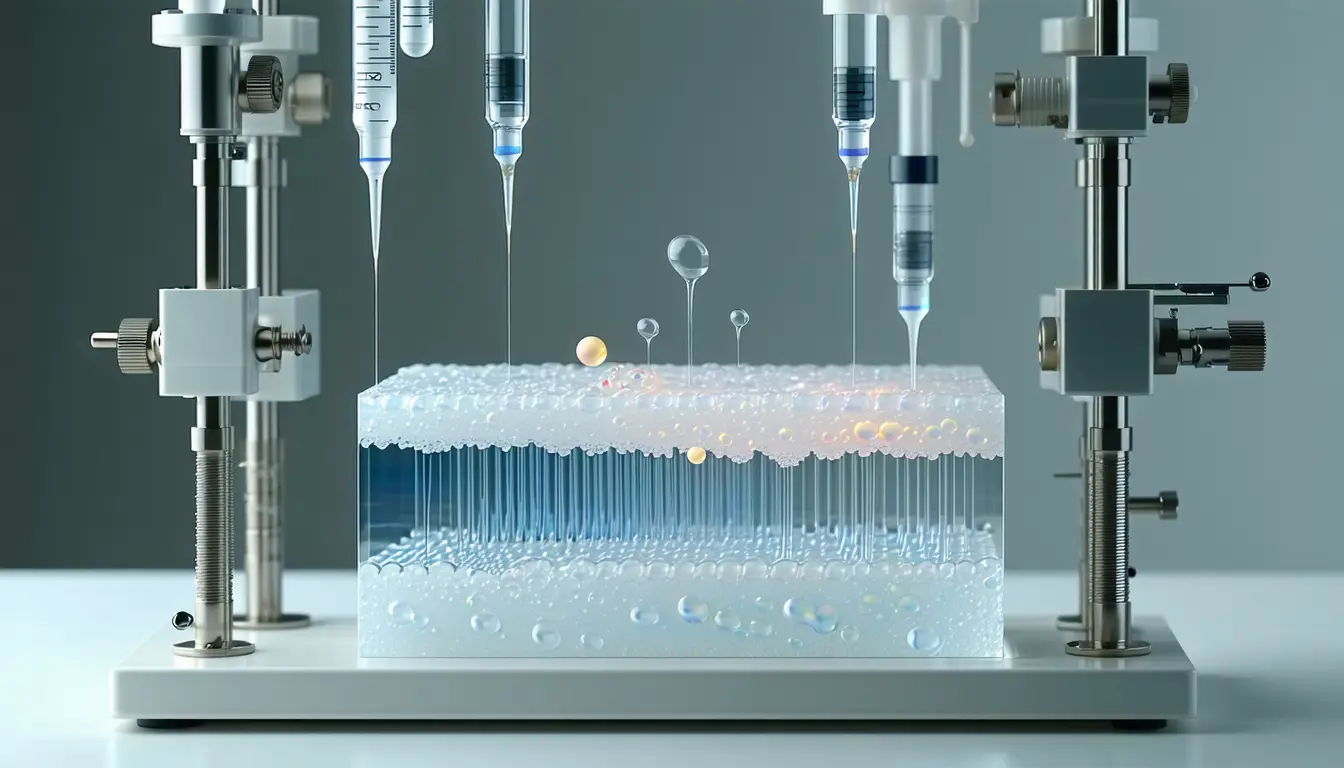
- Researchers at DTU, the University of Copenhagen and University College London built a flexible implant, the microfluidic Axialtrode, with working points spaced along the fiber's length for recording and drug delivery.
- The optical fibers labs use for optogenetics emit light and touch tissue only at the distal tip, which limits an experiment to monitoring or stimulating one brain layer at a time.
- The new fiber starts as a much larger polymer rod that is heated and drawn out, and the finished strand measures less than half a millimetre across.
- A light-conducting core runs down the middle, ringed by eight microscopic channels that can carry liquids or hold extremely thin metal wires for measuring electrical activity.
- The team tested the device in mice and published the work in the journal Advanced Science.
Compiled by The ScientistSomething wrong?How this is made
Why it matters
- capability One insertion can address several depths in the same animal, so a question about signalling between cortical layers no longer needs a separate device per layer.
- constraint Electrodes and drug lines compete for the same eight openings, so how many wires an experiment gets is decided before the animal is on the table.
- decision Anyone treating this as an epilepsy device is reading past the evidence on offer, which is a mouse study of a research instrument.
Each of the eight channels can be a drug line or a sleeve for a recording wire, and the release describes both uses for the same openings [6]. Fluid ports and electrodes therefore come out of one pool: at most eight between them [16]. The release does not say how the team divided the eight in the mouse experiments, or how long the fiber stayed in place [18].
The argument for the design is partly about what the surgery itself does. Kunyang Sui, the postdoc who developed the concept with Associate Professor Christos Markos, said: "Most current brain implants are based on hard materials such as silicon, which can irritate the brain and trigger inflammatory reactions in the tissue. The new implant differs in that it is made of soft, plastic-like optical fibers and has a specially angled tip that makes it smaller and reduces the damage caused when it is placed in the brain" [10][12].
Those are two separable claims. An angled tip is about insertion; softness is about residence, since the fiber bends with tissue instead of pressing rigidly through it, and stiffer implants can provoke inflammation while they sit there [8][17]. The mouse tests reported here speak to whether the fiber functions [4]. What a month in place does to the surrounding tissue is a separate measurement.
For now the group presents the mAxialtrode as a research tool, pointed at how signals travel between brain layers during epilepsy, memory and decision-making [13]. Further out, they describe delivering a drug at one location while stimulating selected areas electrically or with light [14]. Sui cautions that routine clinical use would require extensive testing, further development and regulatory approvals [11].
For a lab that currently implants a probe, a cannula and a light fiber separately, adoption turns on two numbers rather than the count of functions: how well a thin metal wire threaded down a microfluidic channel records, and whether a channel that has held a wire still passes fluid. The work is published in Advanced Science, with DTU, the University of Copenhagen and University College London among the institutions involved [3][2].
What to watch
- Whether the Advanced Science paper reports recording quality and channel patency after weeks in place, not just at insertion.
- Whether a group outside the DTU, Copenhagen and UCL collaboration can draw the fiber and reproduce multi-depth recording and dosing.
- Whether a fiber tuned to bend with mouse tissue can reach deeper structures in larger brains without stiffening.