Science1 publisher3 min readPublished
Replacing the electromagnetic motor with a vibrating glass tube gets an OCT probe to 0.55 mm
A Nanjing group swapped the electromagnetic motor for a piezoelectric crystal and a grooved glass tube, took the imaging probe down to 0.55 mm with one degree of angular error, and threaded it through a full-scale vascular model to the middle cerebral artery.
The Scientist · Science desk
What happened
- Intravascular OCT catheters in clinical use are about 2 mm across and image best in vessels around 10 mm wide. That confines the technique to medium and large arteries.
- A group led from Nanjing has built a probe 0.55 mm across that keeps a full 360 degree field of view and is optimised for vessels about 2 mm wide.
- The probe was pushed through a full-scale human vascular model as far as the middle cerebral artery, the test the team treats as a milestone for cerebrovascular suitability.
Compiled by The ScientistSomething wrong?How this is made
Why it matters
- capability If the probe works in patients, cross-sectional wall imaging becomes available in arteries too narrow and too curved for present catheters, and plaque assessment and stent placement can be planned there.
- constraint The probe takes up 27.5 per cent of its target lumen's diameter against 20 per cent for the conventional pairing, so clearance in tortuous vessels is tighter than the fivefold drop in target vessel size implies.
- decision Anyone weighing the cerebrovascular prospect is deciding on bench, ex vivo and phantom evidence, because no living animal has been imaged with this probe.
- precedent A torsional piezoelectric drive gives catheter builders a route past the electromagnetic micromotor. The micromotor set the floor on catheter diameter.
An electromagnetic motor small enough to spin an OCT lens still needs its windings and its wires, and together they set the floor on catheter diameter at roughly 2 mm [4][3]. The design from Dawei Wu's group at Nanjing University of Aeronautics and Astronautics, with Rui Liu at Nanjing University Medical School, replaces that motor with a piezoelectric one [6]. A crystal on a single-phase AC circuit expands and contracts, vibrating a glass tube along its length. A 10 degree groove in the tube converts the longitudinal vibration into torsion, so the lens traces an ellipse the way a crank and slider does [8]. One phase means very little wiring [10].
From 2 mm to 0.55 mm is a 3.6-fold cut in diameter and about a 13-fold cut in cross-sectional area [1][2]. The optimal target vessel drops from 10 mm to 2 mm [3][7]. Divide probe by vessel and the geometry is less generous than the shrink suggests: the conventional pairing puts a probe across 20 per cent of its target lumen's diameter, the new one across 27.5 per cent [3].
Rotational fidelity is the other half of the problem. A proximal catheter is turned from outside the body, and friction along a curved vessel makes the tip lag and surge, which in traditional proximal catheters shows up as 9 degrees of angular deviation [5][9]. Distal designs move the motor to the tip and pay for it with wires that blank part of the circle [5]. Scanning metal tubes arranged around a small curved vessel, first author Boquan Wang and colleagues held the microprobe to 1 degree at 50 revolutions per second [9], a ninefold improvement and one full cross-section every 20 ms [5][4].
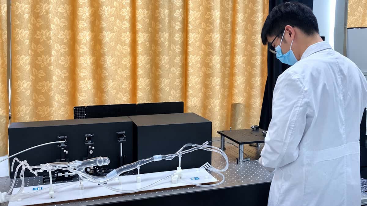
Validation ran in two tracks. Navigation was tested on leaf microveins, a vascular stent and ex vivo pig vessels, then on a full-scale human vascular model, through which the probe reached the middle cerebral artery [11][12]. Lesion detection was tested separately, on human plaque specimens whose OCT images were compared against histology, and the two agreed on where plaques had ruptured and where collagen fibres were dense [13]. None of the tests reported was done in a living animal [6].
So the cerebrovascular result is a navigation result in a phantom. It shows the probe is thin and flexible enough for that path [12]. Whether it still holds 1 degree of angular deviation in a vessel that is moving and pulsing is the next measurement.
Cardiovascular disease is the leading cause of death globally, and atherosclerotic disease accounts for around three-quarters of those deaths [1]. The nearer-term payoff physicsworld describes is narrower: high-resolution images of arterial wall structure in smaller and more curved vessels than clinical catheters can reach today. Plaque assessment and stent positioning would open up in those vessels [14]. The brain's vessels are named as a prospect [15].
What to watch
- An in vivo test: whether the probe holds 1 degree of angular deviation inside a pulsing, moving vessel rather than a bench phantom.
- A published resolution and penetration-depth figure for the 0.55 mm probe: physicsworld reports neither.
- Whether the 50 revolutions per second scan rate is sustained while the probe is pulled back through a small, tortuous vessel.