Science1 publisher2 min readPublished
A swept light sheet records zebrafish seizures starting in the hindbrain and running forward
A University of Georgia microscope imaged zebrafish brains four volumes a second and saw seizures begin at the back and travel to the front, agreeing with one earlier 3D study and disagreeing with two 2D ones. Peter Kner says the field needs more imaging.
The Scientist · Science desk
What happened
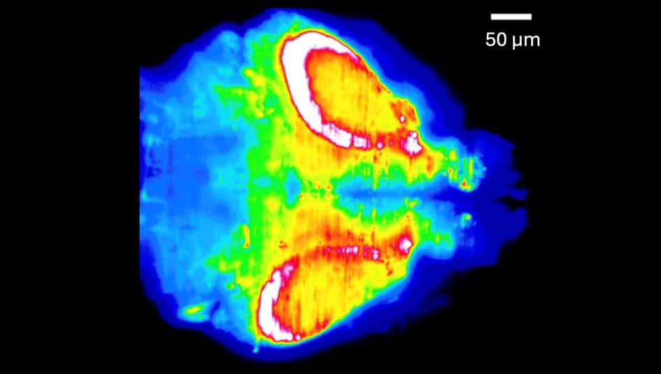
- Engineers at the University of Georgia built a light-sheet microscope for fast volumetric imaging of zebrafish brains and reported the seizure results in Biomedical Optics Express.
- The instrument imaged blocks of tissue measuring 499 by 499 by 148 micrometres at a rate of four volumes a second.
- In a continuous 2.5 minute recording, seizures moved from the back of the fish's brain to the front and then subsided over tens of seconds.
- That hindbrain-first direction matches one earlier 3D imaging study and contradicts two 2D studies published in Frontiers in Neural Circuits and eNeuro.
- Correcting the tunable lens with the pre-calibrated mirror widened the area that stayed near the diffraction limit by a factor of five compared with uncorrected images.
Compiled by The ScientistSomething wrong?How this is made
Why it matters
- capability A 250 millisecond sampling interval puts whole-volume onset ordering inside one seizure, so a lab no longer has to infer sequence by pooling planes recorded from different animals at different times.
- contradiction The volumetric studies agree with each other and the single-plane studies agree with each other, so a reader who consults either literature alone comes away confident and pointed the opposite way.
- constraint With four separate experiments and a two-two split, nothing in the record isolates dimensionality as the cause of the disagreement, and no meta-analysis of the four can fix that.
- decision Groups specifying optics for fast volumetric dynamics now have a concrete choice to make about pre-calibrating aberration corrections per depth, which You expects to carry over to other tunable-lens scanning systems.
Four volumes a second works out to 250 milliseconds a volume [18]. Across the 2.5 minute recording that comes to roughly 600 volumes of the same block of tissue [19], and the seizures took tens of seconds to fade [14]. At that sampling interval, the order in which regions become active can be read inside a single event instead of assembled from separate animals.
Getting there was an optics problem. A light sheet illuminates one plane at a time, which is what keeps background fluorescence from the rest of the sample out of the image [6]. Sweeping that plane through the fish means the collection optics have to follow it, and the group did that with an electrically tunable lens, whose focal length changes with the current through it [7]. Changing that focal length also introduces aberrations, so the researchers measured the correction needed at each depth in advance and held a deformable mirror in the matching shape as the sheet swept [8].
Sixian You, an electrical engineering professor at MIT who was not involved in the work, told Physics World: "This work adds pragmatic utility to 3D light-sheet microscopy for fast volumetric dynamics" [11]. She said deformable mirrors have been used to correct tunable-lens aberrations before, and that the pre-calibration is what makes the fast, wide field-of-view imaging possible [12]. "I expect the same approach will transfer readily to other scanning modalities that stand to benefit from tunable lenses," she said [13].
The direction finding leaves the published record split two against two, with the two volumetric studies on one side and the two single-plane studies on the other [20]. Method and answer line up, across four separate experiments. That correlation between how the imaging was done and which way the wave appeared to go falls short of showing that imaging one plane reverses the apparent direction, and the four papers differ in more than their dimensionality.
Kner, who led the work, is not treating his own result as the settlement. "I think the discrepancies about the direction of seizure propagation point to the need for more imaging," he said [4]. "I believe we still don't know how probabilistic the behaviour is and what factors are important," he said [5].
The report does not give the number of fish imaged or the number of seizure events behind the direction claim [21]. Without those counts, there is no rate to compare against the single-plane literature. The authors put their result as one more data point toward the statistics of seizure propagation, which they say could aid epilepsy treatment if better understood [15].
What to watch
- Kner's team plans to image different zebrafish strains next; if direction varies by strain, dimensionality drops down the list of explanations.
- Whether any group images the same fish in one plane and in volume, which is the direct test of whether single-plane recording reverses apparent direction.
- Whether the pre-calibrated mirror recipe shows up in other tunable-lens scanning systems, as You expects it will.