Science1 publisher3 min readPublished
A cloud-linked miniscope imaged freely moving mice for more than 24 hours straight
Johns Hopkins researchers ran a miniature brain microscope autonomously through a full day of mouse behavior, recording neuronal activity, blood flow and oxygenation, and catching spontaneous seizures hours after a drug dose.
The Scientist · Science desk
What happened
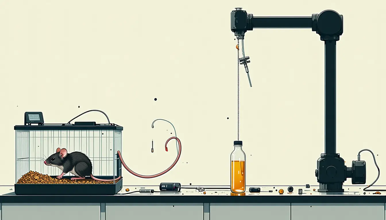
- Johns Hopkins Medicine researchers demonstrated CloudScope, a cloud-based miniature microscope that runs autonomously and images the brains of freely moving mice continuously for more than 24 hours.
- Over those extended runs the device recorded brain activity, blood flow, blood vessel remodeling, oxygenation and cellular behavior, with the data captured and analyzed remotely.
- Pairing the 24-hour imaging record with video of the animals, the team trained an AI framework to say whether a mouse was minimally mobile, moderately active or running from neuronal activity alone.
Compiled by The ScientistSomething wrong?How this is made
Why it matters
- capability A disease-progression study can now be built around events whose timing nobody controls, because the observation period no longer has to be chosen in advance and defended as representative.
- decision Groups planning longitudinal CNS work have to decide whether repeated anesthetized snapshots of separate cohorts still make sense when one animal can be followed continuously and one rig time-shared between labs.
- constraint Johns Hopkins did not report an animal count or a classifier accuracy in the announcement, so a lab cannot yet size a continuous study or judge how much of the behavioral decoding beats guessing.
- exposure Live imaging readable from anywhere puts the data path, and whoever runs the cloud service, inside the experiment's chain of custody.
The design problem is timing. A spontaneous seizure, or the hour a tumor cell starts moving, does not arrive during the two hours an anesthetized animal is under a bench microscope, so a short window samples the disease wherever the experimenter happened to point it. Janaka Senarathna, an assistant professor of radiology at Johns Hopkins, said: "Most central nervous system diseases develop over hours, days or even weeks. Yet modern imaging tools are designed to continuously probe only a small fraction of this time window." [8] He added: "We developed a device to break this time barrier." [9]
The seizure result carries the most weight. The group recorded spontaneous seizures several hours after a drug-induced seizure, and describes them as events conventional short-term imaging would have missed [5]. The strength of that claim comes from the structure of the two methods rather than from a head-to-head measurement in the same animals. It is also the right first result for a continuous instrument: an event a short window cannot see for structural reasons.
Everything here is mouse. Mouse seizure models, and in a separate set of experiments, mouse brain tumors, where the team followed individual cancer cells and changes in the surrounding microenvironment as the disease advanced [6].
The artificial intelligence piece is thinner. Pairing the 24-hour imaging record with video of the animals, the team trained a framework to sort behavior into three classes, minimally mobile, moderately active or running, from neuronal activity alone [7]. Johns Hopkins did not disclose the classifier's accuracy or the number of animals imaged [15]. With three evenly represented classes, a guess lands near 33 percent [14]. Any reported accuracy has to beat that.
Autonomy and remote access are the parts that change how a study is staffed. Live data can be read from anywhere in the world [12], and the architecture supports time-shared imaging, which the group says creates a path toward using fewer animals [11]. If the instrument runs unattended, the biology sets the length of an observation period. The report gives no numbers on throughput or cost.
Arvind Pathak, a professor of radiology, oncology, and biomedical and electrical engineering at Johns Hopkins, said the project started from one question: "We started with a fundamental question: If we wanted to image a seizure or brain tumor formation continuously in a preclinical or animal model over 24 hours or longer, how would we do that?" [10] The work appears in Nature Methods as "A cloud-based miniscope for neurosurveillance of brain health and disease in freely behaving animals" [2]. Next, the team plans to image larger regions of the brain and to use AI to speed up imaging and cancer cell tracking [13].
What to watch
- Whether the Nature Methods paper reports classifier accuracy, animal counts and per-animal validation for the behavior decoding.
- Whether a second lab runs a time-shared CloudScope across sites and reports how many animals a continuous design actually replaces.
- The promised expansion to larger imaging fields and AI-accelerated cancer cell tracking. That expansion would test whether the 24-hour window holds at wider coverage.