Science1 publisher2 min readPublished
Real-time imaging catches CaMKIIα forming chains under synapse-like crowding
A Kanazawa-led team used high-speed atomic force microscopy to watch CaMKIIα holoenzymes stay apart in dilute solution and link into chains once they were packed and confined, with activation widening the spacing by about four nanometres.
The Scientist · Science desk
What happened
- Researchers at Kanazawa University's Nano Life Science Institute, Kyoto University, SOKENDAI and the National Institute for Physiological Sciences report in Science Advances how CaMKIIα organises into chain-like structures.
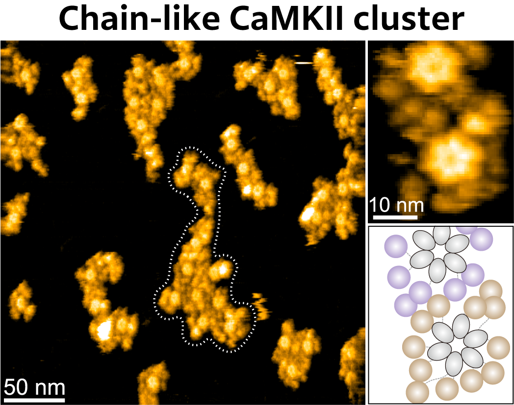
- Under high molecular density and restricted movement, the ring-shaped holoenzymes contacted one another through their catalytic kinase domains and held together as stable chains of several holoenzymes.
- The team also reports that a mutation associated with neurodevelopmental disorders changes how these structures are organised.
Compiled by The ScientistSomething wrong?How this is made
Why it matters
- capability Synaptic CaMKII work has been able to count how much protein gathers at active connections without seeing how the molecules join; single-molecule imaging under crowding gives the joining step itself a direct readout.
- constraint An assay run in dilute solution scores activated and inactive CaMKIIα the same way, so any experiment that skips confinement cannot detect the assembly behaviour this study describes.
- precedent If chain organisation can be scored on a microscope, variant work in neurodevelopmental disorders gains a structural phenotype it can measure directly instead of inferring from kinase activity.
In dilute solution, more than 95 percent of the holoenzymes stayed as single particles, activated or not, and no stable clusters appeared [4]. Fewer than one in twenty were in contact with anything [5]. Switching the protein on did not make it assemble; density and restricted movement did, the two features the team copied from the postsynaptic environment [6].
The contacts sat on the kinase domains, which are the catalytic part of the enzyme [7]. Each holoenzyme is a ring of typically 12 subunits [3], so three linked rings bring 36 kinase subunits into one structure [19].
Calmodulin binding moves those domains outward [9]. In the images, the spacing between neighbouring molecules rose by about four nanometres as the chains got longer, which the authors take as confirmation that the activated protein had opened into a more extended shape [10]. A phosphate added at one autophosphorylation site kept the larger chains stable [11], and the simulations reproduced the same pattern: opened molecules with restricted movement are more likely to form bigger groups [12].
"Our observations connect the structural changes of individual CaMKIIα holoenzymes with their collective organization at a larger scale," Shibata said [13]. "The results suggest that activation does more than switch on kinase activity: it also changes how CaMKIIα molecules assemble with one another" [14].
Clusters began forming at densities below those estimated inside the postsynaptic density, the protein-rich signalling region of a synapse [8]. The comparison carries the biological claim, and the density it is measured against is an estimate. No measurement of that density was made in the same experiment.
The disease half of the study is thinner in the published account, which says a mutation associated with neurodevelopmental disorders changes how the structures are organised without identifying the variant or the size of the change [20].
Whether the chains form in a living spine is still an open question. The protein was purified and the crowding was built by the experimenters [6]. Nothing here was measured in a neuron or about memory. The team's account of long-term potentiation, in which activated holoenzymes dock on receptors at the receiving side of a connection and then seed chains around themselves, is a proposal [16] about one of the main routes to memory formation [17].
What to watch
- Whether the same chains appear in intact dendritic spines, by cryo-electron tomography or imaging in live neurons.
- The identity of the neurodevelopmental-disorder variant and how far it shifts chain length or contact geometry.
- A mutation that blocks the kinase-domain contact surface while leaving catalysis intact; it would separate assembly from kinase activity.