Science1 publisher2 min readPublished
Human heart muscle cells divide after a heart attack in tissue taken during bypass surgery
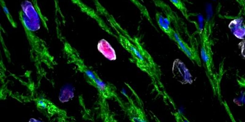
A University of Sydney group sampled living hearts during bypass operations and found human heart muscle cells dividing after a heart attack, the response mice were known to mount and no one had shown in people.
The Scientist · Science desk
What happened
- Researchers at the University of Sydney, the Baird Institute and Royal Prince Alfred Hospital report in Circulation Research that the human heart produces new muscle cells after a heart attack, against the assumption that it cannot.
- Increased mitosis in heart muscle after an infarct had been seen in mice; this is the first demonstration of the same phenomenon in humans.
- The team says the natural response is not currently strong enough to replace all the muscle a heart attack destroys.
Compiled by The ScientistSomething wrong?How this is made
Why it matters
- capability Compounds that raised heart muscle cell division in mice now have living human tissue to be tested in, in a laboratory model that may track what happens in patients more closely than post-mortem material.
- constraint Supply of that tissue depends on cardiac surgery patients consenting to a sample mid-operation, which caps how large any human study of this process can get and how fast it can run.
- contradiction The announcement is framed around a heart that can regrow muscle, while its first author says the regrowth is not enough to prevent a heart attack's effects; the second reading governs how far this sits from a therapy.
Mitosis is a cell caught in the act of dividing. Counting it tells you a division started. It does not tell you that a working muscle cell finished the job. The University of Sydney announcement carries no counts at all: not the number of dividing cells, not the number of patients sampled [17]. Those are the figures an amplification strategy needs, because a drug that doubles a rate is only interesting once you know what it is doubling. A heart attack can destroy as many as one third of the cells in the human heart [4].
The sampling is what lifts this above an autopsy observation. Diseased and non-diseased tissue came out of the same heart, so each patient supplies their own comparison and differences between donors drop out of it [6]. The technique was developed by Paul Bannon and Sean Lal, who hold joint appointments at the University of Sydney, Royal Prince Alfred Hospital and the Baird Institute [7]. Bypass patients are a particular population: chronic coronary disease, an elective operating date. The material does not say how long after their heart attacks the tissue was taken [17], and that interval decides whether the response is a brief window after injury or something still running in scarred muscle years later.
Robert Hume, the paper's first author and lead of translational research at the Baird Institute [10], was direct about the distance between the finding and a treatment. "Although this new discovery of regrowing muscle cells is exciting, it isn't enough to prevent the devastating effects of a heart attack. Therefore, in time, we hope to develop therapies that can amplify the heart's natural ability to produce new cells and regenerate the heart after an attack," he said [8].
The alternative explains why a weak repair process is worth chasing at all. Australia has about 144,000 people living with heart failure and performs about 115 heart transplants a year [13][14]. Divide one by the other and you get roughly 1,252 people with the condition for every transplant performed [15]. Transplant is the only cure the University of Sydney names [12], and cardiovascular disease accounts for nearly 24 percent of Australian deaths [3].
What to watch
- The Circulation Research paper's own figures: how many patients, and what fraction of heart muscle cells were in division in diseased versus non-diseased regions.
- Whether follow-up work shows the dividing cells complete division into functional muscle rather than arresting partway.
- Whether compounds that raised heart muscle cell division in mice get run through the living human tissue model at Royal Prince Alfred.