Science1 publisher2 min readPublished
Patterned light writes each cell's position into a barcode that single-cell sequencing can read
scSTAMP-seq uses patterned light and photocleavable barcodes to record each cell's position before standard single-cell multiome sequencing. So far it has been tested only in human embryoids, and labs will need resolution and error figures before they can weigh it against a dedicated spatial platform.
The Scientist · Science desk
What happened
- Researchers described scSTAMP-seq, a light-encoded barcoding method designed to keep spatial information while running on standard single-cell platforms.
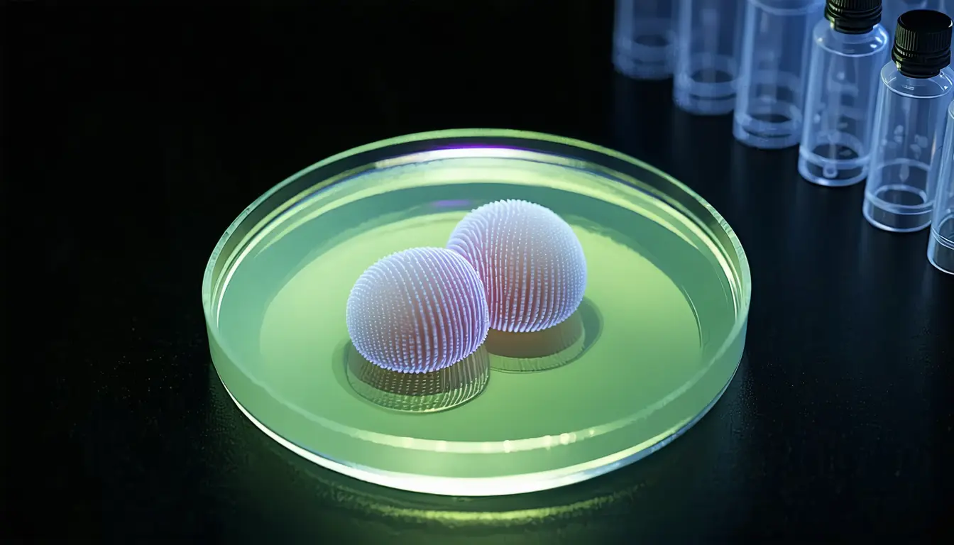
- Applied to human embryoids, the method is reported to show spatial organization and epigenetic states jointly regulating cellular programs.
- Raw and processed sequencing data are public in NCBI's Gene Expression Omnibus under accession GSE237524.
Compiled by The ScientistSomething wrong?How this is made
Why it matters
- capability A lab already running single-cell multiome capture could add position to its data by changing sample preparation and illumination, keeping the sequencing platform it already has.
- constraint Human embryoids are the only system reported, so use on tissue sections or clinical samples is an extrapolation until someone tests it.
- decision Groups weighing the method can re-examine its position calls in the deposited data before changing their own protocols.
Standard single-cell sequencing is good at saying what a cell is and poor at saying where it was. According to the paper's abstract, scSTAMP-seq is built to keep the second answer while still running on standard single-cell platforms [1].
The protocol has two steps before the cells reach a sequencer. First, live or fixed cells are labelled with hashtag oligonucleotides that carry a cholesterol conjugate and can be cut by light [2]. Then patterned light is projected onto the sample. In the abstract's word, that exposure 'stamps' spatial coordinates onto individual cells before single-cell multiome sequencing [3].
The paper's title calls the patterns "topological light gradients" and names transcriptomics and epigenomics as the readouts [5]. A cell's position ends up recorded in the state of its photocleavable tag, and the sequencing run reads that tag along with the rest of the cell's data [8].
I think this is the right place to put the spatial step. Position is written in chemistry on the cell, so the capture instrument never has to handle coordinates [1][3]. The extra work moves to sample preparation and to whatever projects the light.
That view holds only if the numbers do. The abstract does not report a spatial resolution, a cell count, a rate of cells assigned to the wrong position, or the illumination hardware used [9]. Those figures decide how the method compares with dedicated spatial methods such as Slide-seq and Slide-seqV2, both of which appear in the paper's reference list [7].
The biological claim needs the same care. According to the abstract, the human embryoid data show "how spatial organization and epigenetic states coregulate cellular programs" [4]. Measuring a cell's position and its epigenetic state in the same experiment can show that the two co-vary. To establish which one sets the other, you need an experiment that changes one and watches what happens to the other.
What to watch
- Figures from the full paper on spatial resolution and on how often cells are assigned to the wrong position.
- An independent reanalysis of GSE237524, or a replication in tissue sections by another group.
- Whether the photocleavable hashtag reagents and a light-patterning setup become available off the shelf, and what they cost next to a dedicated spatial platform.