Science1 distinct publisher2 min readPublished Updated
In 56 patients and 57 matched controls, a technique called SANDI produced the pattern post-mortem studies describe, and only in the regions the disease attacks. Making it a trial endpoint needs repeat scans of the same people.
The Scientist · Science desk

Compiled by The ScientistSomething wrong?How this is made
The thalamus is the part of this design worth admiring. Compare only the basal ganglia between two groups and any measurement that happens to differ for unrelated reasons will look like disease. Including a structure that Huntington's largely spares in early stages makes the study its own control, and the pattern duly appeared in the basal ganglia while the thalamus stayed clean [9][10].
Direction matters as much as magnitude. Apparent cell-body density fell, apparent cell-body size rose, and the space between cells widened [10]. One number moving down would be weak evidence; two moving in opposite directions is a signature, and it is the signature pathologists have described in tissue, with a particular class of striatal neurons lost while glial cells enlarge and become more active [11]. These are model outputs, inferred from how water moves through tissue rather than counted cells [6].
Age sits inside the headline number. In some striatal regions, the apparent cell-body estimates together with a participant's age explained up to 63% of the observed shrinkage [13]. The account the authors published does not separate what the imaging adds from what age alone would have delivered. "Up to" also picks out the best-performing regions, so even there roughly 37% of the shrinkage goes unexplained [17].
The denominator is 113 people scanned once, 56 with Huntington's and 57 volunteers matched for age and sex [7][16], and the authors call the study a snapshot [14]. Every association it reports therefore runs across people rather than within one, including the links to disease severity and to finger-tapping performance [12]. On the published evidence this is a cross-sectional marker of striatal cellular change, which is a real thing to have and a different thing from a measure that follows one patient's biology as it changes.
What the scan does not measure is whether shifting these numbers helps anyone move or think better. About 8,000 people in the UK live with Huntington's, there is no cure, and the cell and gene therapies that would need such a readout are still being tested [1][2][3]. The authors put it carefully: SANDI might, in future trials, help determine whether a treatment is altering the underlying disease process, and they say they are not quite there yet [15].
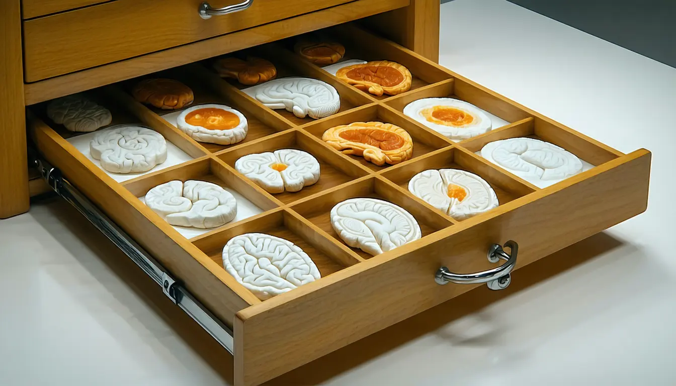
Ranked by verification strength, evidence, and original report placement.
About 8,000 people in the UK are living with Huntington's disease, an inherited condition that gradually affects movement, thinking and mood.
Huntington's disease is caused by a faulty gene, its effects usually begin between the ages of 30 and 50, and there is currently no cure.
New cell and gene therapies for Huntington's disease are being developed and tested.
One of the main things that happens in Huntington's is loss of neuronal cells in the striatum, part of the basal ganglia, and as cells are lost the brain tissue in these regions shrinks.
Conventional MRI can show the shrinkage but a standard brain scan tells relatively little about what is happening inside the tissue at a cellular level.
The team used soma and neurite density imaging (SANDI) to analyse diffusion MRI, which detects how water moves through brain tissue and so allows indirect estimates of properties such as the apparent size and density of cell bodies.
Distinct publishers with included, body-backed reporting in this cluster.
1 article · September 5, 2026
Follow any of these and your For You feed starts watching them — no settings page required.
science
Feral hogs are now a budget problem: containment is buyable, eradication is not1 distinct publisher
invest
Alaska's disappearing lakes make permafrost thaw an asset-level observable1 distinct publisher
science
HIPAA Covers Less Than You Think, And "Anonymized" Is Not A Legal Shield1 distinct publisher
science
MRI catches structural brain change seven years before amyloid-PET sees plaque1 distinct publisher
Evidence-backed comparisons of source perspectives and observed adoption signals. Read the methodology
Which Builder, Operator, and Investor concerns the observed source mix emphasized—not a truth score.
Evidence, demonstrated adoption, hype gap, incentives, and confidence are assessed independently, each on its own current evidence. How these are measured.
Well-designed, singly-reported
The design carries real weight: 113 people, controls matched on age and sex, and a region the disease spares early used as an internal check that the signal is not a generic artefact. Concordance with what autopsies describe, selective neuron loss beside enlarged glia, is the kind of external anchor a new imaging measure needs. Against that, everything arrives through the researchers' own summary, with no journal named, no intervals on the correlations, and no one outside the team reading the scans.
No use beyond the study
Nothing in this reporting shows the technique in use anywhere outside the study. The scans came off a strong-gradient scanner and the authors say the method must still be adapted and validated for the machines hospitals actually run; no trial, sponsor or imaging site is named as taking it up. Scoring uptake here would mean inventing it.
Headline ahead of a snapshot
"Could help treat Huntington's disease" promises more than a single-timepoint comparison can deliver; what was shown is that a diffusion measure separates patients from controls in the right regions. The gap stays modest because the authors close it themselves, spelling out that they cannot yet say whether SANDI tracks progression or registers a drug effect.
The method's authors hold the pen
This is a researcher-written column about the researcher's own technique, and the future it sketches, SANDI as a readout in cell and gene therapy trials, is also the future that funds the work. Scientific American adds its own subscription pitch partway down. None of that makes the findings wrong; it does mean no party in this story had reason to look for the result's weak spots.
One consistent account, unchecked
The internal numbers hang together and the reported limitations match the design, which is why this sits at the midpoint rather than lower. It does not sit higher because a single self-authored account is the whole record: no second outlet, no named journal, and no independent expert to say whether the biophysical model's assumptions hold in tissue that is losing neurons and gaining reactive glia at the same time.