Science1 publisher2 min readPublished
A five-minute per-patient fit puts MIT's X-ray-to-CT registration under a millimeter
Existing 2D/3D registration models align well for some patients and fail for others. MIT's xvr, out today in Nature, fits itself to one patient in about five minutes, then matches that patient's X-rays to their 3D scan in seconds.
The Scientist · Science desk
What happened
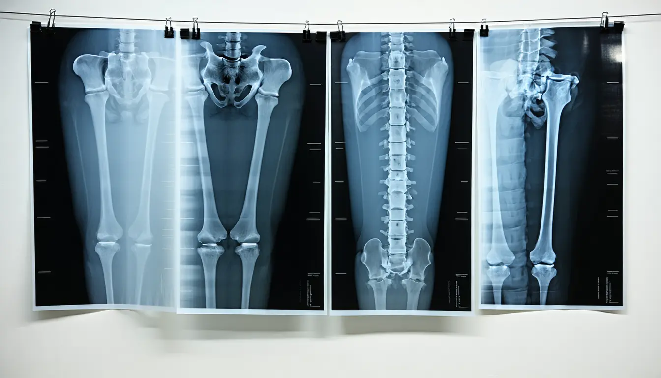
- Scientists and clinicians at MIT and collaborating institutions published xvr in Nature, a system for matching X-rays taken during a procedure to the patient's preoperative CT or MRI.
- MIT reports that xvr outperformed existing AI methods by an order of magnitude across a wide range of patients, body parts, and medical procedures.
- Earlier AI registration tools align images well for some patients and fail for others; the MIT release says that has made them infeasible in practice.
Compiled by The ScientistSomething wrong?How this is made
Why it matters
- capability Second-scale alignment at sub-millimeter precision is fast enough to tell a clinician where a catheter sits in three dimensions while it is still moving.
- constraint Five minutes of fitting has to fit somewhere in the schedule, and the stroke intervention Gopalakrishnan cites is the case with the least room for it.
- decision A hospital deciding whether to trial this has image-space error to weigh and no patient outcomes, so the first decision is whether to run that study itself.
- precedent Fitting a model to a single patient at use time is an answer to the label shortage that constrains medical imaging models generally, and other groups facing the same shortage now have a worked example.
A model trained once and shipped to every hospital has to cope with anatomy it has never seen. The existing tools break there, according to the MIT account: they align images well for some patients and fail for others. That inconsistency has kept them out of practice [5]. xvr treats the individual as the training target. It spends about five minutes adapting to one patient [2], then matches that patient's X-rays to their 3D scan in a matter of seconds, with sub-millimeter precision [3].
The reason to build it that way is data. A network robust enough to handle many patients needs high-quality annotated medical images, and there are not enough of them, according to Gopalakrishnan [10]. Per-patient fitting works around the shortage, because the one high-quality volume you can count on is the patient's own preoperative CT or MRI, which is the scan clinicians are aligning to anyway [13].
MIT says xvr beat existing AI methods by an order of magnitude across a wide range of patients, body parts, and procedures [4]. MIT did not disclose the metric behind that comparison, complication rates, or procedure times [16]. If the comparison is on registration error, then ten times sub-millimeter puts the methods being compared at millimeter scale or worse [15].
Gopalakrishnan put the clinical case in terms of travel time. "A majority of Americans live more than an hour away from a center that can perform noninvasive procedures, like emergency stroke interventions. An hour in stroke time is incredibly substantial," he said [8].
What the system is replacing is either a person's trained intuition or a slow manual workaround. "It takes decades of training for a clinician to become skilled enough to see grainy, 2D images and understand how everything is oriented," Gopalakrishnan said [9]. The manual alternative has the clinician guess an instrument's position by punching numbers into a computer or clicking anatomical landmarks on a screen [6].
Sub-millimeter registration is a measurement on images. Whether it shortens a thrombectomy or lowers the complication rate that flat X-rays contribute to [7] is a measurement on patients, and only a prospective study in a working suite produces it. Two co-authors work in those rooms: Andrew Abumoussa, a neurosurgeon at St. Luke's Marion Bloch Neuroscience Institute, and Darren B. Orbach, a physician and scientist at Boston Children's Hospital [12].
What to watch
- Whether the Nature paper reports per-case failures on unseen anatomy, and across how many patients.
- Whether the roughly five-minute fit can run on the preoperative scan ahead of time, because an emergency stroke case does not have five minutes to spare.
- A prospective report of registration error measured during live procedures, with complication rates alongside it.