Science1 distinct publisher3 min readPublished
A Morgridge Institute team read metabolism in unlabeled immune cells and left the sample usable afterwards, which makes cell-therapy manufacturing the likelier first customer, though nobody has yet tied the signal to a product outcome.
The Scientist · Science desk

Compiled by The ScientistSomething wrong?How this is made
The contrast comes from the cells themselves. A sensitive microscope delivers two long-wavelength photons, which do not damage material the way shorter wavelengths do, and the readout depends on intrinsic fluorescence rather than anything the operator adds [16] [6]. What the published account does not give is the part an engineer would want: no classification accuracy, no list of which cells were confused for which, no throughput figure.
The sample matters more than the laser. Earlier ways to measure immune cell metabolism required isolating one cell type at a time or adding chemical labels that mark metabolites [10], and standard flow cytometry, which is the workhorse here, does not routinely assess metabolism at all even though metabolic state reveals subpopulations and functional states that surface markers miss [9]. Riendeau and colleagues worked in the whole mononuclear mixture, which the team argues gives more accurate insight into behaviour than a monoculture would [15]. It is also the harder problem, since PBMCs span T, B and NK lymphocytes plus monocytes [17], and separating which cell a signal came from is entangled with what state that cell is in.
The denominator is worth stating plainly. The images came from undifferentiated PBMCs isolated from the donated blood of three human volunteers [5], so between-person variation is being estimated from three points, or two degrees of freedom [18]. The study shows single-cell metabolic phenotyping works inside a complex sample, but three donors cannot support a reference range. No patient samples appear in this account: the blood cancers, lupus, sepsis and cognitive decline in the framing are stated as hopes for the technique [13], not cohorts it was tested against.
The manufacturing use is the more interesting one, because reagent-based metabolic assays can harm the cells they measure [8], and Riendeau's claim is that the analysis leaves the sample intact and usable afterwards [7]. In a cell-therapy workflow, a destructive assay is an assay you pay for twice, once in reagents and once in cells you can no longer infuse. PBMCs are the starting material for CAR T therapies [2], and clinical labs currently report the abundance of cell types with some basic functional measures [1]. Skala's own framing is conditional: a lot of starting material for cell therapies is PBMCs, and it would be useful to assess the fitness of those cells before processing them [11].
What is missing is a link between a day-zero metabolic signature and anything a manufacturer is scored on. Nothing here links autofluorescence lifetimes to expansion, transduction efficiency, product potency or patient response. The authors' own conclusion is that optical metabolic imaging could complement traditional PBMC measurements and improve disease monitoring and immune therapy development [14]. Complement is the correct verb. A research capability becomes a release test only when someone shows the number moves with an outcome, and then sets a limit on it.</body_markdown> </invoke>
Ranked by verification strength, evidence, and original report placement.
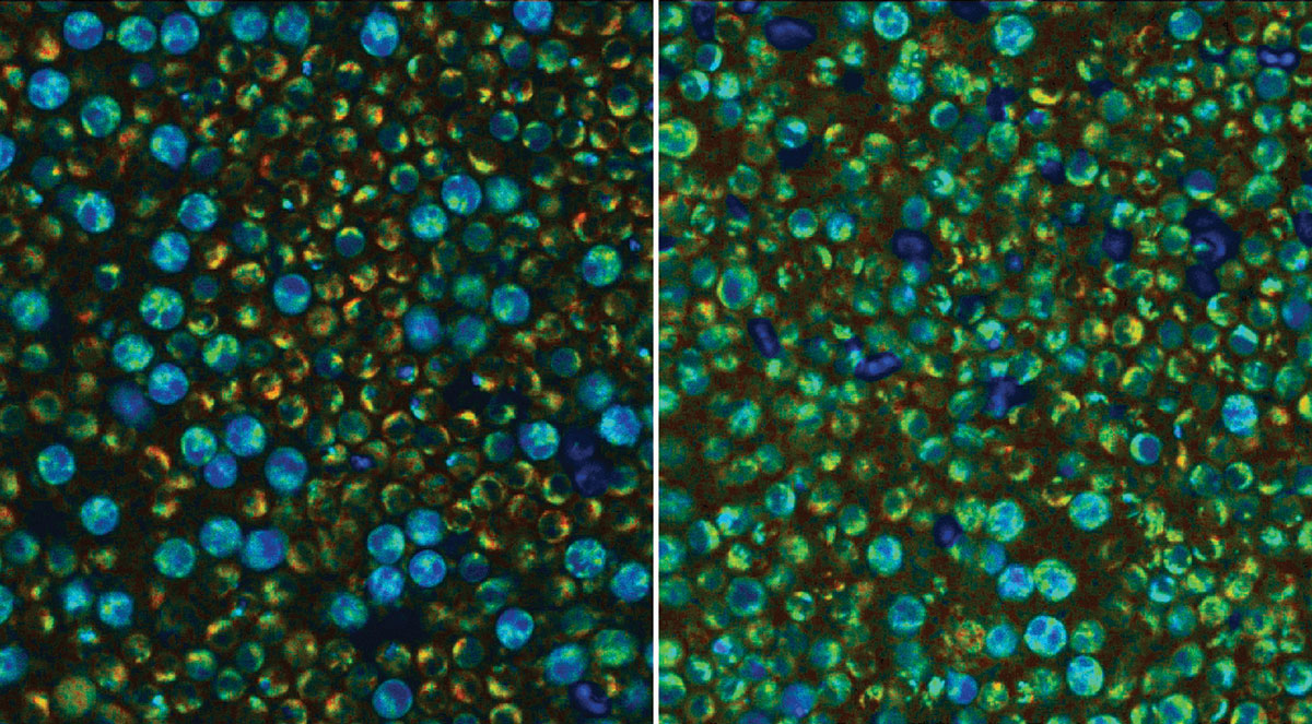
A study by scientists at the Morgridge Institute for Research demonstrated that optical metabolic imaging (OMI) can be used to characterize metabolic activity within immune cells from the peripheral blood of patients.
First author Jeremiah Riendeau says the team has for the first time accurately observed the metabolic state of single immune cells across a complex PBMC sample using a nondestructive analysis, and that the samples can continue to be used afterwards.
Riendeau says that with other techniques, studying metabolism requires adding different reagents to the sample, and that this can be harmful to the cells.
Previously, techniques to measure immune cell metabolism needed to isolate individual cell types or add chemical labels that highlight the presence or absence of metabolites.
Clinical labs isolate peripheral blood mononuclear cells (PBMCs) from a blood draw and assess the abundance of different cell types along with some basic measures of how they are functioning.
PBMCs are the starting point for CAR T cell therapies, which engineer a patient's own immune cells to fight certain cancers.
Distinct publishers with included, body-backed reporting in this cluster.
1 article · September 1, 2026
Follow any of these and your For You feed starts watching them — no settings page required.
science
Salk's fly driver lines narrow octopamine access to single pairs of neurons1 distinct publisher
science
267 Ubiquitin Receptors, 93 Already Diseased: A Catalog That Gives Variants a Mechanism1 distinct publisher
science
Hollings researchers turn chemoresistance into a target by blocking LOX first1 distinct publisher
science
Chemically tuned suppressor tRNAs read through CFTR's premature stop in mice and patient organoids1 distinct publisher
Evidence-backed comparisons of source perspectives and observed adoption signals. Read the methodology
Which Builder, Operator, and Investor concerns the observed source mix emphasized—not a truth score.
Evidence, demonstrated adoption, hype gap, incentives, and confidence are assessed independently, each on its own current evidence. How these are measured.
Well quoted, unreplicated
Every figure and quote reaches the reader through one trade outlet's reading of one paper — but GEN quotes that paper directly, names both authors, and publishes the accuracy spread rather than only the novelty claim, including the 74% on NK cells that cuts against the pitch. That is careful reporting of a result nobody outside the authoring lab has yet touched: three donors, one journal, no outside confirmation.
Still inside the originating lab
Nothing has been taken up. The imaging method was pioneered in Skala's own lab and applied to blood from three volunteers; no other group is named running it, no manufacturer is trialling it on feedstock, and no patient sample has been read for a clinical decision. The closest thing to uptake here is the paper itself.
One step ahead of the data
The paper's own verbs are cautious — 'could provide', 'complement' — and GEN largely keeps them. The stretch happens around the edges: a three-donor demonstration of subset and activation calls is set beside blood cancers, lupus, sepsis and cognitive decline, and 'for the first time' does heavy lifting for a claim only its authors have made. Overstated by a step rather than a leap.
Inventors as sole witnesses
The technique's developers are also its only witnesses here. Skala's lab pioneered the imaging approach, the case for its clinical worth is made in Skala's and Riendeau's quotes about their own paper, and it is relayed by a biotech trade outlet whose readership is the intended customer base. No rival method's advocate, no independent clinician and no would-be buyer appears in the account.
Firm on the what, thin on the so-what
Who did it, where it ran, what was measured and how accurately are all solid enough to quote back. The forward half — that this reading of metabolism tells a manufacturer or a clinician something they would act on — rests on hope, and with two degrees of freedom for donor variation there is very little here to generalise from.